Iliopsoas fascia (Latin: fascia iliopsoas)
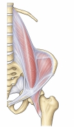
· The iliopsoas fascia is the fascia that covers the front of the iliopsoas (psoas major, iliacus and, when present, psoas minor). It is made up of the part over psoas major (the psoas fascia) and the part over iliacus (the iliac fascia), which are continuous with each other.
· Many textbooks describe this whole fascia under the name iliac fascia, as "the fascia covering the front of the inner hip muscles (iliopsoas)" (see Iliac fascia).
Position and continuity
· The fascia of psoas major is thin above (cranially) and thicker below (caudally), where it joins the fascia of iliacus. Above, it is thickened into the medial arcuate ligament of the diaphragm.
· Laterally it is attached to the inner lip of the iliac crest; medially to the bodies of the lumbar vertebrae and, in the pelvis, to the periosteum along the pelvic brim (iliopectineal line) of the lesser pelvis, where it blends with the pelvic fascia.
· Below, it is fixed firmly to the back of the inguinal ligament and is continuous with the transversalis fascia. It then passes into the thigh with iliopsoas and covers the muscle down to its insertion on the lesser trochanter of the femur; it also forms the lateral wall of the femoral sheath. At its lateral edge, beside quadratus lumborum, it is continuous with the fascia over the front of that muscle (the anterior layer of the thoracolumbar fascia).
· Older textbooks put it this way: the psoas fascia, thin above and thick below, joins the iliacus fascia, is firmly bound to the lateral part of the inguinal ligament, and covers iliopsoas as a strong connective tissue membrane down to its insertion.
Iliopectineal arch, muscular and vascular lacunae
· Between the inguinal ligament and the iliopubic eminence the fascia is thickened into a band, the iliopectineal arch. It divides the space under the inguinal ligament into the lateral muscular lacuna (for iliopsoas and the femoral nerve) and the medial vascular lacuna (for the femoral artery and femoral vein).
· The femoral nerve and the lateral femoral cutaneous nerve run deep to this fascia, between the fascia and iliopsoas.
· A psoas abscess can spread down inside the sheath formed by this fascia and appear as a swelling in the groin or the upper thigh. In anaesthesia, local anaesthetic is injected deep to the fascia to block the femoral nerve and its neighbours together (the fascia iliaca block).
More detail
· See the pages Iliac fascia, Iliopectineal arch, Muscular lacuna and Iliopsoas.
Reference websites
[Wikipedia]
· Iliac fascia, Wikipedia.
· Iliopsoas, Wikipedia.
Related terms
- Body of ilium
- Ala of ilium
- Iliac tuberosity
- Iliopubic eminence
- Iliopectineal fossa
- Iliac fascia
- Iliopsoas
- Psoas major
- Iliacus
- Fascia
- Thoracolumbar fascia
- Inguinal ligament
- Vascular lacuna
References and sources
- Further reading: Iliac fascia, Wikipedia.
- Further reading: Iliopsoas, Wikipedia.
- Federative International Programme for Anatomical Terminology (FIPAT). Terminologia Anatomica, 2nd ed. (TA2), 2019.