Jugular fossa of temporal bone (Latin: fossa jugularis ossis temporalis)
· On the lateral side of the fossa a narrow groove for the mastoid canaliculus soon enters the bone as the mastoid canaliculus, which runs backwards and opens at the tympanomastoid fissure; it carries the auricular branch of the vagus nerve. The small triangular depression in front of the fossa, along the posterior border of the petrous part, is the external aperture of the cochlear canaliculus.
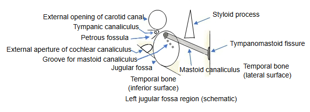
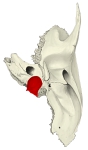 Left temporal bone (inferior view)
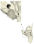 Left temporal bone (around the jugular fossa)
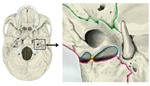 External cranial base: left jugular foramen
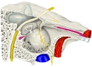 Right tympanic cavity (oblique sagittal section)
 Dural venous sinuses
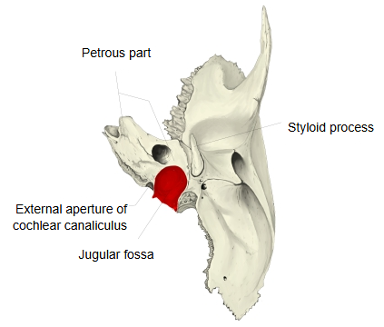
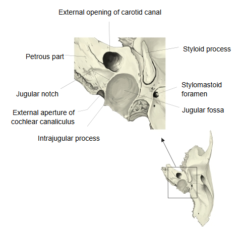
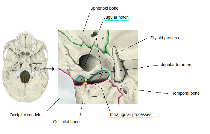
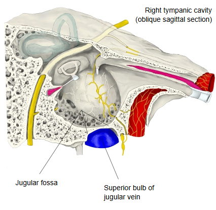
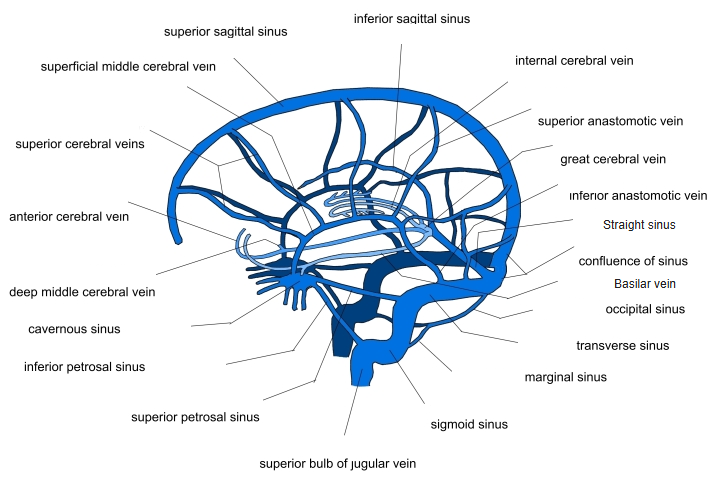
Near the middle of the posterior border, on the inferior surface of the petrous part, lies the large, deep, arched jugular fossa. It holds the superior bulb of the jugular vein, and its roof separates the bulb from the floor of the tympanic cavity.
Reference websites
[Wikipedia]
· Jugular fossa, Wikipedia.
[Websites with illustrations]
· Left temporal bone, inferior surface (Gray's Anatomy, plate 141) (Wikimedia Commons)

· Base of the skull, outer surface (Gray's Anatomy, plate 187) (Wikimedia Commons)

· The temporal bone: parts and landmarks (TeachMeAnatomy)
The fossa lies behind the carotid canal and the external aperture of the cochlear canaliculus. On the bony ridge between the carotid canal and the fossa is the inferior opening of the tympanic canaliculus, which transmits the tympanic nerve, a branch of the glossopharyngeal nerve. Behind the fossa a four-sided rough jugular surface, covered with cartilage in life, joins the jugular process of the occipital bone. The depth and size of the fossa vary between skulls and between the two sides.
Related terms
- Jugular foramen
- Jugular notch
- Carotid canal
- External aperture of cochlear canaliculus
- Groove for mastoid canaliculus
- Petrous part
References and sources
- Further reading: Jugular fossa, Wikipedia.
- Federative International Programme for Anatomical Terminology (FIPAT). Terminologia Anatomica, 2nd ed. (TA2), 2019.