Paranasal sinuses (Latin: sinus paranasales)
What are the paranasal sinuses?
Specifically, there are the following four.
sinuses
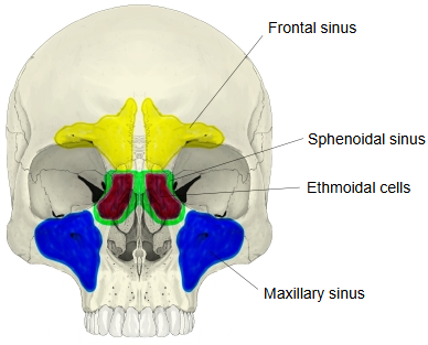
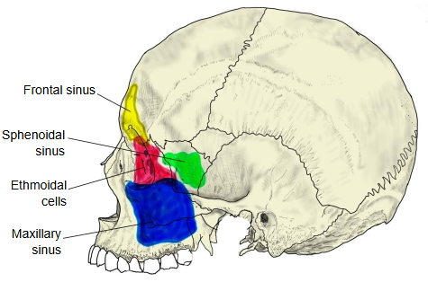
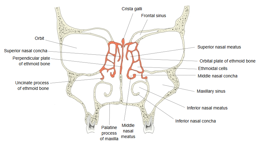
- Maxillary sinus : in the body of the maxilla; the largest paranasal sinus. Opens into the middle nasal meatus.
- Frontal sinus : in the frontal bone, behind the superciliary arches. Opens into the middle nasal meatus.
- Ethmoidal cells : in the ethmoid, divided into many small air cells between the nasal cavity and the orbit. The anterior and middle cells open into the middle meatus, the posterior cells into the superior meatus.
- Sphenoidal sinus : in the body of the sphenoid, lying behind the ethmoidal cells. Opens into the sphenoethmoidal recess.
Why do they exist?
Their function is not certain; the following roles have been suggested.
1. Making the skull lighter
2. Adding resonance to the voice
3. Buffering the temperature difference between the outside air and the skull
Development and clinical notes
The maxillary sinuses and ethmoidal cells are present at birth but small; the frontal and sphenoidal sinuses appear in childhood. All grow considerably around puberty.
Infection can spread to them from the nose (sinusitis). The maxillary sinus drains poorly because its opening is high on its medial wall.
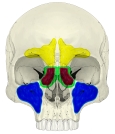 Paranasal sinuses (anterior)
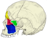 Paranasal sinuses (left lateral)
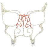 Nasal cavity and sinuses (coronal section)
Reference websites
[Wikipedia]
· Paranasal sinuses, Wikipedia.
[Websites with illustrations]
· Position of the paranasal sinuses in the head (Wikimedia Commons)

· Paranasal sinuses and the ostiomeatal complex (side and front views) (aboutcancer.com)

· The paranasal sinuses (TeachMeAnatomy)
Related terms
- Maxillary sinus
- Frontal sinus
- Ethmoidal air cells
- Sphenoidal sinus
- Nasal cavity
- Middle nasal meatus
- Sphenoethmoidal recess
References and sources
- Further reading: Paranasal sinuses, Wikipedia.
- Federative International Programme for Anatomical Terminology (FIPAT). Terminologia Anatomica, 2nd ed. (TA2), 2019.