Posterior ethmoidal foramen (Latin: Foramen ethmoidale posterius)
Note: some sources, such as English Wikipedia, place the opening of the posterior ethmoidal foramen a little further back than described above, close to the sphenoid. It usually lies about 10–12 mm behind the anterior ethmoidal foramen (see the illustrations below).
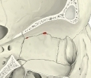 Ethmoidal foramina
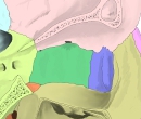 Bones of the medial orbital wall
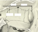 Distances between the foramina
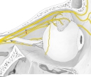 Orbit (lateral view, section)
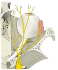 Orbit (superior view)
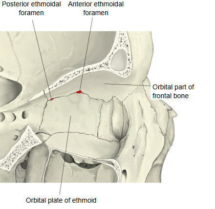
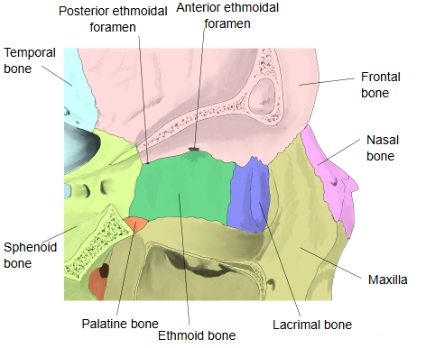
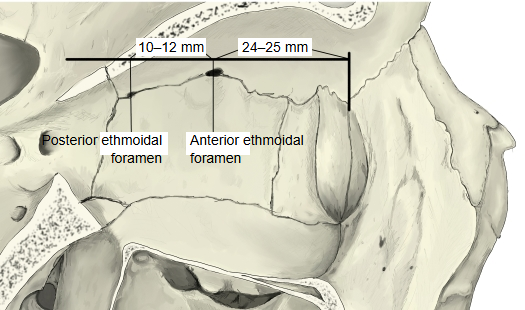
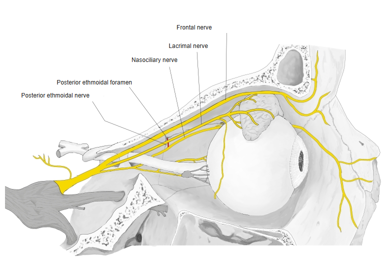
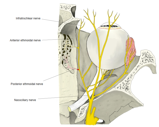
The posterior ethmoidal foramen is the posterior one of the two ethmoidal foramina. It lies between the upper border of the orbital plate of the ethmoid and the orbital part of the frontal bone, behind the anterior ethmoidal foramen.
It is the passage for the posterior ethmoidal nerve and the posterior ethmoidal artery and vein, and its canal opens towards the posterior ethmoidal cells of the ethmoidal labyrinth. On the cranial side, its internal opening lies lateral to the olfactory groove, at the side of the cribriform plate.
Reference websites
[Wikipedia]
· Posterior ethmoidal foramen, Wikipedia.
· Ethmoidal foramina, Wikipedia.
[Websites with illustrations]
· Left human orbit, labelled (including an ethmoidal foramen) (Wikimedia Commons)

· Ethmoid bone (Kenhub)
Related terms
- Orbit
- Orbital plate
- Orbital part of frontal bone
- Ethmoidal air cells
- Ethmoidal labyrinth
- Posterior ethmoidal artery
- Posterior ethmoidal nerve
References and sources
- Further reading: Ethmoidal foramina, Wikipedia.
- Federative International Programme for Anatomical Terminology (FIPAT). Terminologia Anatomica, 2nd ed. (TA2), 2019.