Bursae around the knee joint
Also called the bursae of the knee
Note: some of them are not present in every person.
The bursae of the knee are small sacs lined with synovial membrane and filled with synovial fluid; they reduce friction where tendons, ligaments and skin move over bone. Some communicate with the joint cavity (for example the suprapatellar bursa and the popliteus bursa), others are separate. Thin-walled pouches that open into the joint enlarge the joint space but are also weak points of the joint. They can be grouped as communicating and non-communicating bursae or, as below, by position: in front, on the lateral side and on the medial side (including behind the knee). Inflamed bursae (bursitis) are a common cause of local swelling and pain.
Anterior
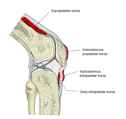
| 1 | Suprapatellar bursa |
| Between the front of the lower end of the femur and the deep surface of the tendon of quadriceps femoris; it lets the tendon slide over the femur and usually opens into the joint cavity. | |
| 2 | Subcutaneous prepatellar bursa |
| Between the patella and the skin; it lets the skin move over the patella. Its inflammation is called "housemaid's knee". | |
| 3 | Deep infrapatellar bursa |
| Between the upper part of the tibia and the patellar ligament; it lets the ligament move over the tibia. | |
| 4 | Subcutaneous infrapatellar bursa |
| Between the patellar ligament and the skin. | |
| 5 | Subcutaneous bursa of tibial tuberosity (pretibial bursa) |
| Between the tibial tuberosity and the skin; it lets the skin move over the tuberosity. |
Lateral
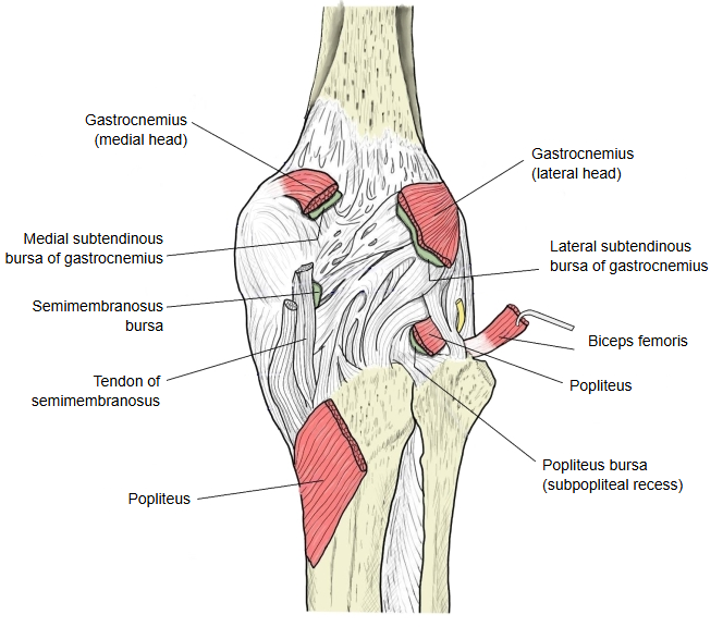
| 1 | Lateral subtendinous bursa of gastrocnemius |
| Between the lateral head of gastrocnemius and the joint capsule. | |
| 2 | Inferior subtendinous bursa of biceps femoris (fibular bursa) |
| Between the fibular (lateral) collateral ligament and the tendon of biceps femoris. | |
| 3 | Fibulopopliteal bursa |
| Between the fibular collateral ligament and the tendon of popliteus. | |
| 4 | Subpopliteal recess (popliteus bursa) |
| Between the tendon of popliteus and the lateral condyle of the femur; an outpouching of the joint cavity. |
Medial
| 1 | Medial subtendinous bursa of gastrocnemius |
| Between the medial head of gastrocnemius and the joint capsule; it often communicates with the joint. | |
| 2 | Anserine bursa |
| Between the tibial (medial) collateral ligament and the tendons of sartorius, gracilis and semitendinosus (the pes anserinus). | |
| 3 | Semimembranosus bursa |
| Between the tibial collateral ligament and the tendon of semimembranosus. | |
| 4 | |
| A bursa between the tendon of semimembranosus and the medial condyle (head) of the tibia. | |
| 5 | |
| Occasionally a bursa between the tendons of semimembranosus and semitendinosus. |
· A swollen bursa behind the knee, usually linked with the medial gastrocnemius and semimembranosus bursae, is called a popliteal (Baker's) cyst.
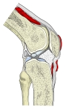 Bursae in front of the knee
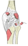 Bursae behind the knee
Reference websites
[Wikipedia]
· Bursae of the knee, Wikipedia.
· Synovial bursa, Wikipedia.
Related terms
- Knee joint
- Patella
- Patellar ligament
- Quadriceps femoris
- Gastrocnemius
- Semimembranosus
- Tibial collateral ligament
References and sources
- Further reading: Bursae of the knee, Wikipedia.
- Further reading: Synovial bursa, Wikipedia.
- Federative International Programme for Anatomical Terminology (FIPAT). Terminologia Anatomica, 2nd ed. (TA2), 2019.