Porta hepatis (Latin: porta hepatis)
Also called the hilum of liver or the hilar fissure or the transverse fissure of liver
· Its approximate size (rough estimates; sources differ):
length (from side to side) about 5–8 cm; height (front to back) about 2–3 cm; depth about 1.5–2 cm. Many textbooks give its length as about 5 cm.
The porta hepatis and the fibrous capsule
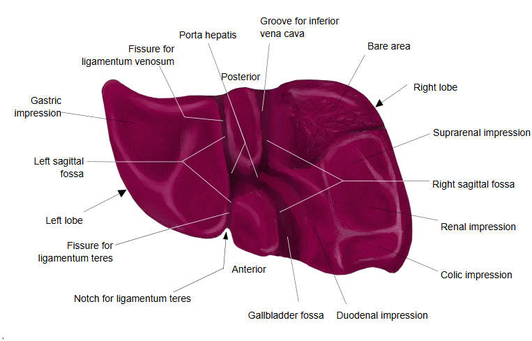
· The porta hepatis is a fissure-like opening on the inferior surface of the liver; its walls are formed by the liver parenchyma itself.
· The opening is not closed over by a separate membrane. Instead, the fibrous capsule of the liver (Glisson's capsule) turns inward at its margins.
· The structures passing through the porta hepatis (the portal vein, the hepatic artery and the bile ducts) enter the liver wrapped in loose connective tissue, which continues from the capsule as the perivascular fibrous sheath (Glisson's sheath).
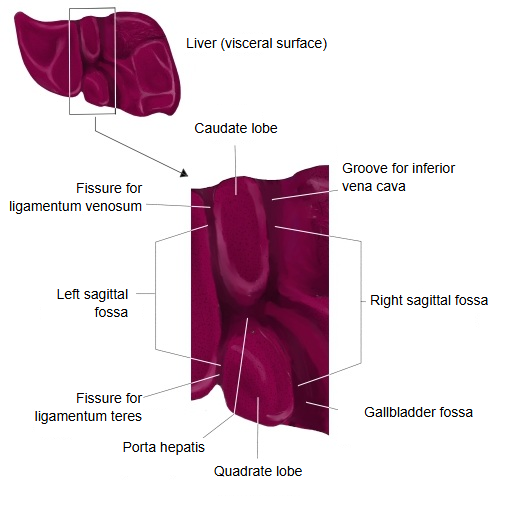
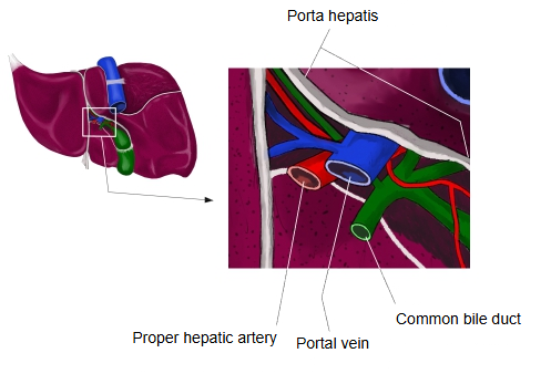
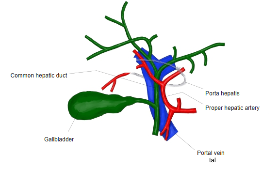
· From front to back, the right and left hepatic ducts (leaving), the branches of the proper hepatic artery (entering) and the branches of the portal vein (entering) pass through the porta hepatis; the ducts lie to the right, the artery to the left and the vein behind them. These structures are often called the portal triad. The fissure for the ligamentum teres meets the left end of the porta hepatis.
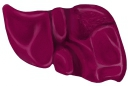 Liver (visceral surface)
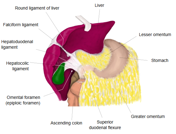
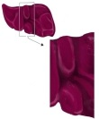 Around the porta hepatis
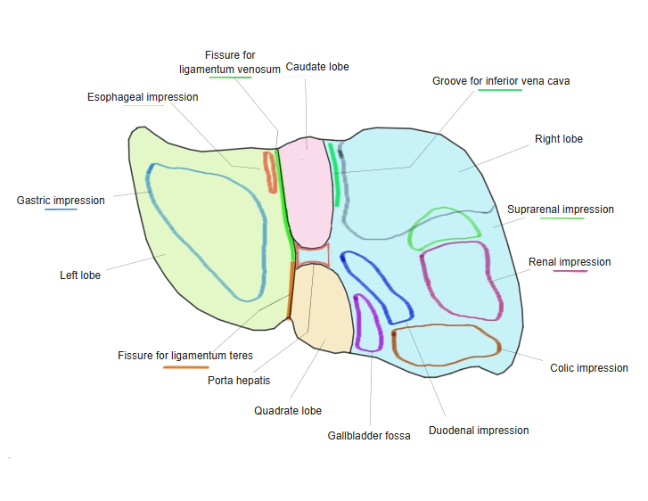
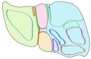 Features of the visceral surface
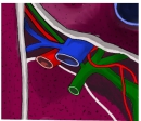 Porta hepatis
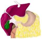 Hepatoduodenal ligament
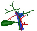 Vessels at the porta hepatis
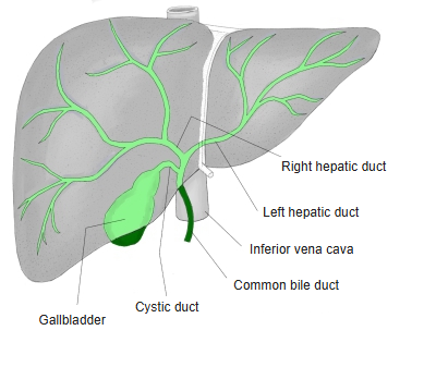
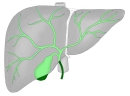 Bile ducts
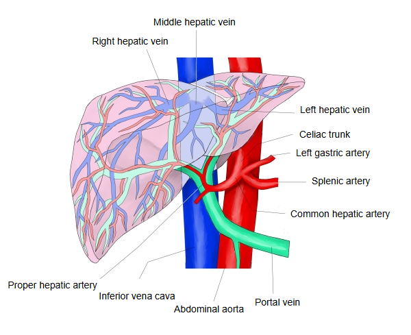
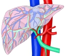 Vessels of the liver
Structures passing through it
| Artery | Proper hepatic artery | A branch of the common hepatic artery; at the porta hepatis it divides into right and left branches, which enter the liver. |
| Vein | Portal vein | Enters the liver and branches; together with the branches of the hepatic artery it opens into the capillaries of the liver (sinusoids). The blood is then gathered again into veins and leaves as the hepatic veins, which drain into the inferior vena cava. The hepatic veins do not pass through the porta hepatis; they leave on the posterior surface of the liver. |
| Bile ducts | Right and left hepatic ducts | The right hepatic duct collects bile from the right lobe and the left hepatic duct from the left lobe. They leave the liver parenchyma at the porta hepatis and unite to form the common hepatic duct, which later joins the cystic duct to form the common bile duct. |
| Nerves | Autonomic nervous system | Sympathetic: nerve fibres from the celiac plexus. Parasympathetic: fibres of the vagus nerve, branching from the anterior and posterior vagal trunks, run to the liver as hepatic branches. Together they form the hepatic plexus. |
| Lymphatic vessels | The lymphatic vessels entering and leaving the liver (superficial and deep lymphatic vessels) are an important drainage route of the liver. The liver produces more lymph than any other organ (often estimated at roughly a quarter to a half of all the lymph in the body). Hepatic lymph nodes lie near the porta hepatis. | |
Reference websites
[Wikipedia]
· Porta hepatis, Wikipedia.
· Liver, Wikipedia.
[Websites with illustrations]
· Porta hepatis (Radiology Key)

· Porta hepatis (illustration) (Pinterest (image))

Where it is found
Related terms
- Portal triad
- Hepatoduodenal ligament
- Caudate lobe
- Quadrate lobe
- Fissure for ligamentum teres
- Common hepatic duct
- Liver
References and sources
- Further reading: Porta hepatis, Wikipedia.
- Further reading: Liver, Wikipedia.
- Federative International Programme for Anatomical Terminology (FIPAT). Terminologia Anatomica, 2nd ed. (TA2), 2019.