Laryngeal muscles (Latin: musculi laryngis)
Textbooks describe them as follows.
The larynx has many small muscles that move it as a whole or move its cartilages against each other. They form two groups: the extrinsic muscles, which attach the larynx (or the hyoid bone) to surrounding structures, and the intrinsic muscles, which run between the laryngeal cartilages.
· Extrinsic laryngeal muscles: change the position of the cartilages, raising and lowering the larynx, for example during swallowing.
· Intrinsic laryngeal muscles: change the shape of the glottis and the length and tension of the vocal folds, so they control breathing, protection of the airway and the voice.
Extrinsic muscles that raise the larynx: thyrohyoid, stylopharyngeus, palatopharyngeus, and the suprahyoid muscles acting through the hyoid bone.
Extrinsic muscles that lower the larynx: sternothyroid, and sternohyoid and omohyoid acting through the hyoid bone (members of the infrahyoid muscles).
Below is a short list of the intrinsic laryngeal muscles, based on a classical anatomy textbook.
laryngeal
muscles
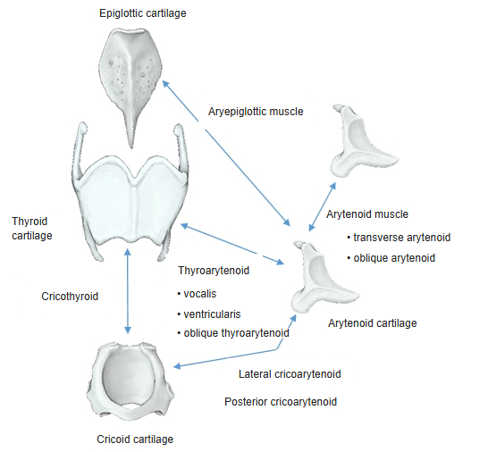
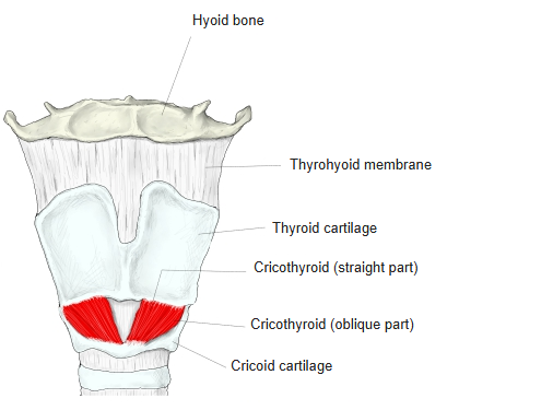
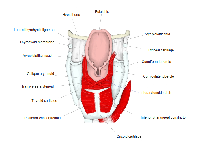
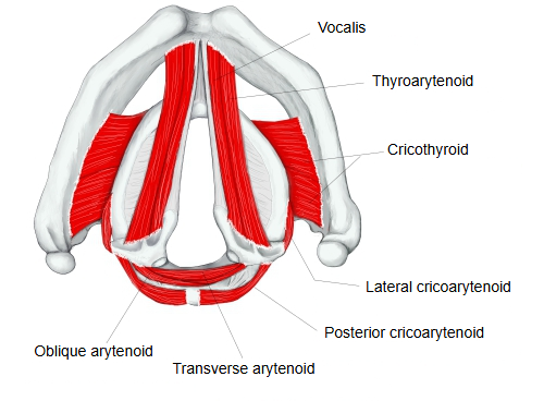
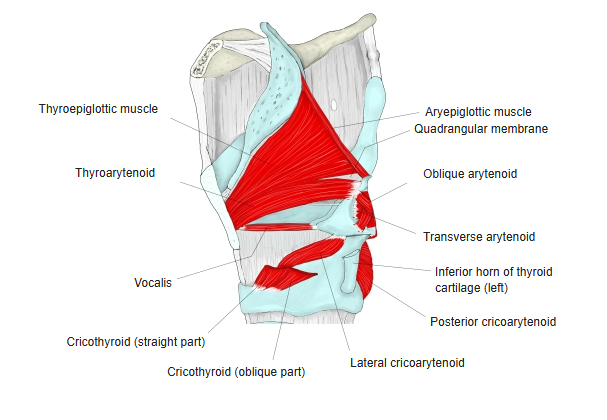
- Cricothyroid : from the outer surface of the arch of the cricoid cartilage to the lower border and inferior horn of the thyroid cartilage. Tilts the thyroid cartilage forwards and so lengthens and tenses the vocal folds.
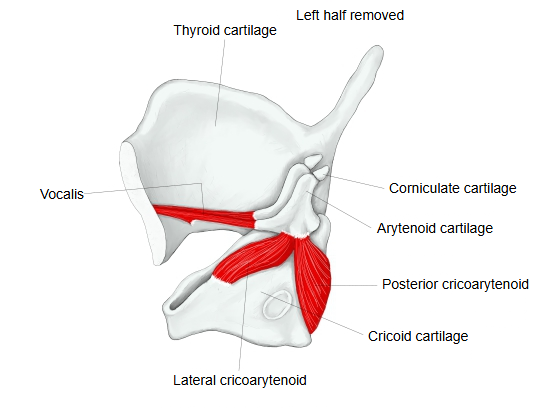
- Posterior cricoarytenoid : from the back of the cricoid lamina to the muscular process of the arytenoid cartilage. The only muscle that opens the rima glottidis.
- Lateral cricoarytenoid : from the upper border and outer surface of the side of the cricoid cartilage to the muscular process and the front of the arytenoid. Closes the front part of the rima glottidis.
- Arytenoid : fills the hollow on the backs of the two arytenoid cartilages and ends on their outer borders; closes the back part of the rima glottidis; two kinds:
- Transverse arytenoid : unpaired; runs horizontally between the outer borders of the two arytenoids.
- Oblique arytenoid : from the muscular process of one arytenoid to the apex of the other.
- Aryepiglottic : from the upper outer surface of the arytenoid to the epiglottic cartilage; usually described as the continuation of the oblique arytenoid in the aryepiglottic fold.
- (External) thyroarytenoid : from the inner surface of the thyroid cartilage to the muscular process and oblong fovea of the arytenoid. Shortens and relaxes the vocal folds. Fibres that reach the epiglottis form the thyroepiglottic part.
- Thyroarytenoid (inner parts) : inside the laryngeal cavity; two parts:
- Vocalis : from the back of the thyroid cartilage near the midline to the vocal process and oblong fovea; lies beside the vocal ligament.
- Ventricular part : from the back of the thyroid cartilage near the midline to near the colliculus of the arytenoid.
- Oblique thyroarytenoid : from the muscular process of the arytenoid to the upper border of the thyroid cartilage.
Texts differ in how finely they divide the arytenoid and thyroarytenoid groups and which parts they name as separate muscles.
Nerve supply: all intrinsic muscles are supplied by the recurrent laryngeal nerve (through the inferior laryngeal nerve), except the cricothyroid, which is supplied by the external branch of the superior laryngeal nerve.
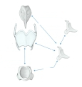 Laryngeal cartilages and muscles
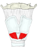 Larynx (anterior)
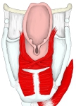 Larynx (posterior)
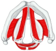 Larynx (superior view)
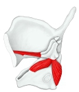 Right half of the larynx (from the left)
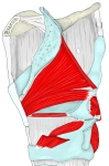 Intrinsic laryngeal muscles
Related terms
- Cricothyroid
- Infrahyoid muscles
- Suprahyoid muscles
- Recurrent laryngeal nerve
- Superior laryngeal nerve
- Vocal fold
- Larynx
References and sources
- Further reading: Larynx, Wikipedia.
- Further reading: Intrinsic muscles of the larynx, Wikipedia.
- Federative International Programme for Anatomical Terminology (FIPAT). Terminologia Anatomica, 2nd ed. (TA2), 2019.