Scala tympani
Also called Tympanic duct
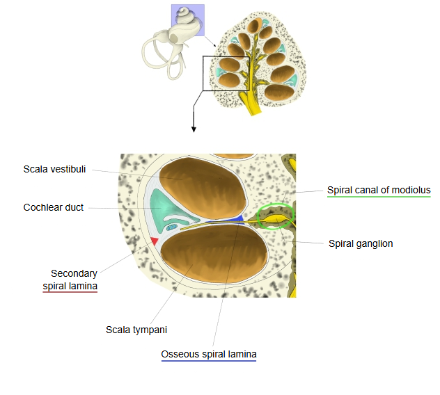
- Spiral canal of the cochlea : total length about 3 cm, about 2.5–2.75 turns.
- Upper level
- Scala vestibuli : filled with perilymph; carries the sound vibrations to the cochlear duct; part of the bony labyrinth. Joined to the scala tympani at the apex through the helicotrema.
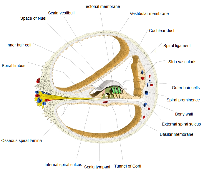
- Cochlear duct : separated from the scala vestibuli by the vestibular (Reissner's) membrane; filled with endolymph and contains the spiral organ (organ of Corti); both ends are blind. Contents: tectorial membrane, organ of Corti, internal and external spiral sulci, spiral prominence, stria vascularis.
- Lower level
- Scala tympani : part of the bony labyrinth, filled with perilymph; passes the travelling wave on to the tympanic cavity. Its boundary with the upper level: the osseous spiral lamina + the basilar membrane (+ the secondary spiral lamina).
- Upper level
- Modiolus : the cone of spongy bone at the centre of the cochlea, with the longitudinal canals and the spiral canal of the modiolus.
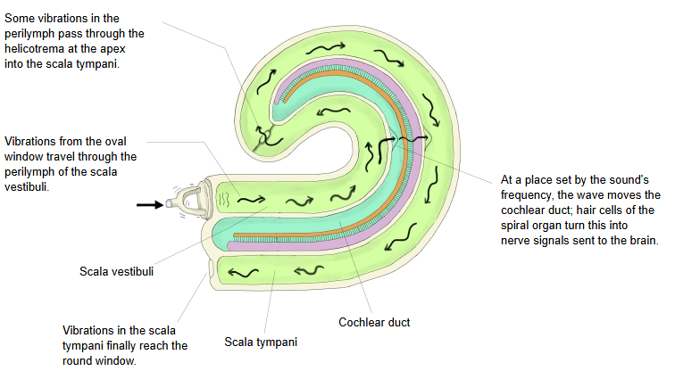
Sound vibrations enter the perilymph of the scala vestibuli at the oval window. The travelling wave they set up runs along the basilar membrane and peaks at a place that depends on the frequency; there the cochlear duct with the vestibular membrane above and the basilar membrane below moves, and the shearing between the tectorial membrane and the hair cells turns the movement into electrical signals.
Not all of the energy that reaches the cochlear duct goes to the hair cells: the pressure wave passes through the basilar membrane into the scala tympani and is released at the round window.
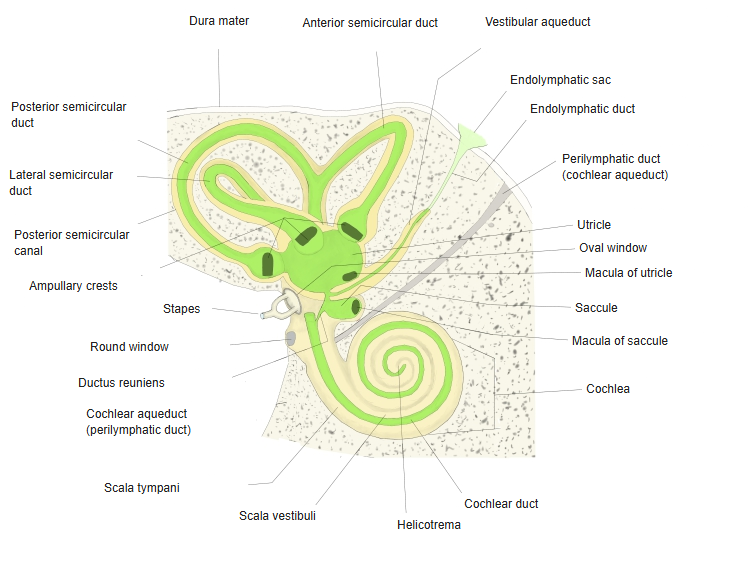
Many illustrations show the path of sound in this way; the figure 'Path of sound in the cochlea' below was drawn on that basis. Near the round window the scala tympani also receives the opening of the cochlear canaliculus (the perilymphatic duct), through which the perilymphatic space communicates with the subarachnoid space.
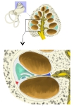 One turn of the cochlea
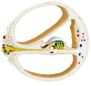 Cochlear duct (cross-section)
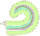 Path of sound in the cochlea
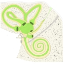 Bony and membranous labyrinths
Reference websites
[Wikipedia]
· Tympanic duct, Wikipedia.
[Websites with illustrations]
· The inner ear (TeachMeAnatomy)
· Longitudinal section of the cochlea (Gray's Anatomy, fig. 928) (Wikimedia Commons)

Related terms
References and sources
- Further reading: Tympanic duct, Wikipedia.
- Federative International Programme for Anatomical Terminology (FIPAT). Terminologia Anatomica, 2nd ed. (TA2), 2019.