Tympanic plexus (Latin: plexus tympanicus)
Nerves that form it
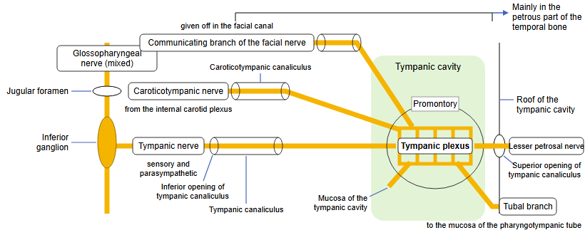
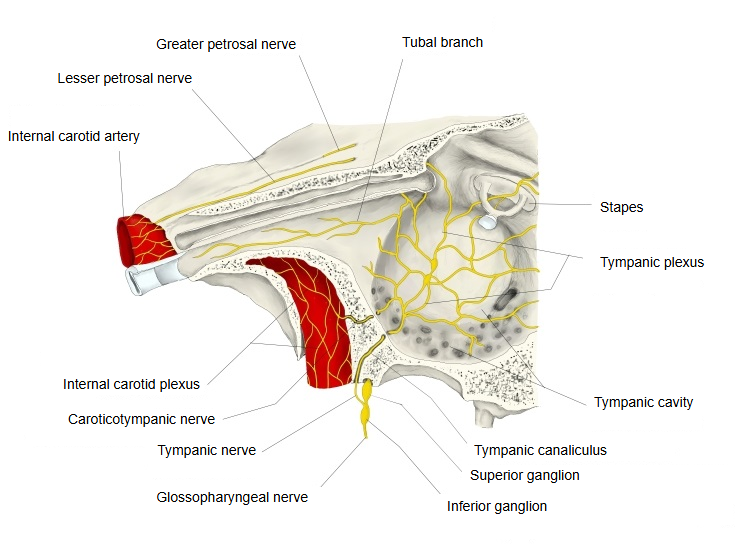
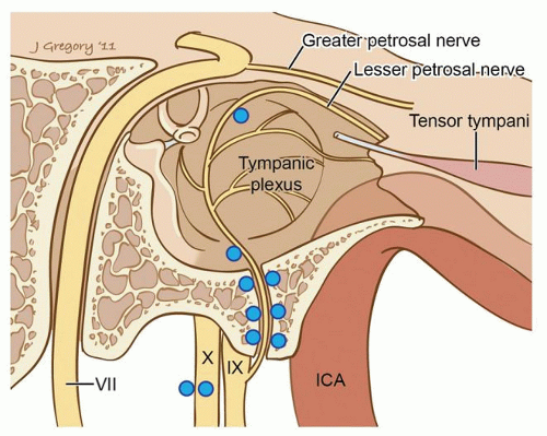
Main root: the tympanic nerve, a branch of the glossopharyngeal nerve that arises from its inferior ganglion and reaches the cavity through the tympanic canaliculus.
Nerves that join it:
1. 1. The communicating branch of the facial nerve: it enters the tympanic cavity through the superior opening of the tympanic canaliculus.
2. 2. The caroticotympanic nerves: sympathetic branches of the internal carotid plexus that enter the tympanic cavity through the caroticotympanic canaliculi.
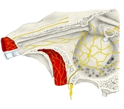 Left tympanic cavity (anterolateral view)
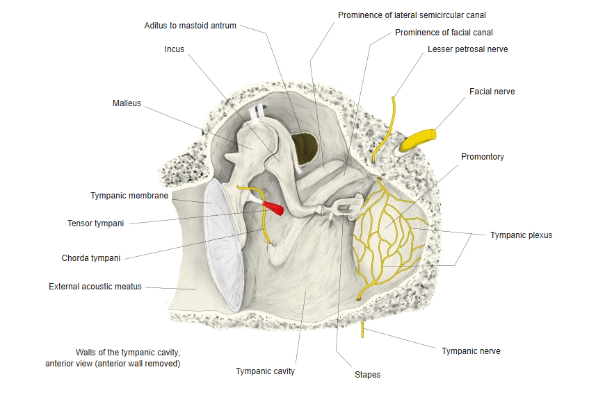
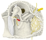 Walls of the left tympanic cavity (anterior view)
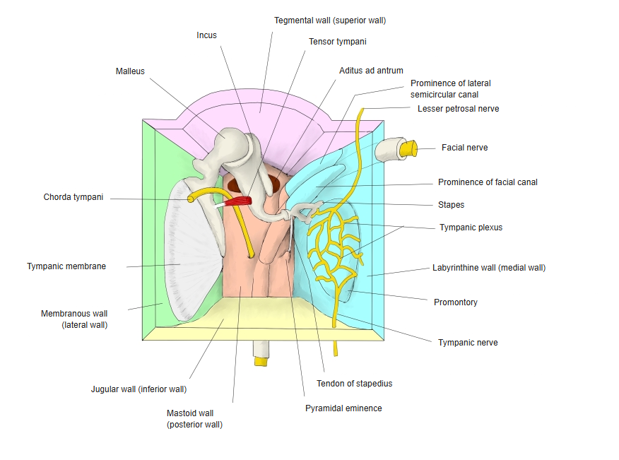
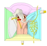 Tympanic cavity (model, anterior view)
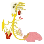 Glossopharyngeal nerve (model)
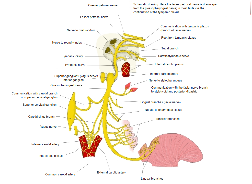
- Superior ganglion : then through the jugular foramen.
- Inferior ganglion : communicates with the auricular branch of the vagus nerve.
- Tympanic nerve : to the tympanic plexus, which communicates with a branch of the facial nerve and with the caroticotympanic nerves; from the plexus: the lesser petrosal nerve and the tubal branch.
- Stylopharyngeal branch
- Pharyngeal branches
- Carotid sinus branch : to the carotid sinus and carotid body.
- Tonsillar branches
- Lingual branches : communicate with the lingual nerve.
Reference websites
[Wikipedia]
· Tympanic plexus, Wikipedia.
[Websites with illustrations]
· Tympanic plexus on the promontory, with the tympanic and caroticotympanic nerves (figure) (doctorlib.info)

· Tympanic plexus and the petrosal nerves (figure) (Entokey)
Related terms
- Superior opening of tympanic canaliculus
- Inferior opening of tympanic canaliculus
- Tympanic nerve
- Glossopharyngeal nerve
- Lesser petrosal nerve
- Caroticotympanic nerve
- Tympanic cavity
- Otic ganglion
- Caroticotympanic canaliculi
References and sources
- Further reading: Tympanic plexus, Wikipedia.
- Federative International Programme for Anatomical Terminology (FIPAT). Terminologia Anatomica, 2nd ed. (TA2), 2019.