Clavipectoral fascia (Latin: fascia clavipectoralis)
Also called: coracoclavicular fascia; costocoracoid membrane (upper part)
One classic description of the deep pectoral fascia reads as follows.
Under the pectoralis major, the deep fascia covers the pectoralis minor and becomes stronger as it passes upwards and laterally, forming the clavipectoral fascia. It is strengthened in its upper part, between the first rib and the coracoid process, where it is called the costocoracoid membrane, and it has openings for vessels and nerves. Above it is attached to the clavicle and the coracoid process, medially to the upper costal cartilages (the fascia over the first intercostal spaces and the first rib), and laterally it fuses with the sheath of the axillary vessels and continues into the axillary fascia. The fascia of the subclavius encloses that muscle and blends with fibres of the coracoclavicular ligament.
In that description the fasciae of the chest are divided into three:
of the
chest
- Pectoral fascia : covers the front surface of the pectoralis major
- Deep pectoral fascia : lies under the pectoralis major and covers the pectoralis minor; its upper part is the clavipectoral fascia
- Endothoracic fascia : lines the whole inner surface of the thoracic cage
Other descriptions name the three layers differently.
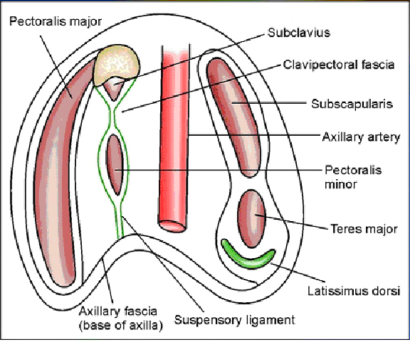
In them the thoracic wall has three fasciae: the superficial pectoral fascia, the clavipectoral fascia and the endothoracic fascia. The clavipectoral (coracoclavipectoral) fascia lies behind the pectoralis major, on the pectoralis minor and the subclavius, and is stronger than the superficial fascia. It is firmly attached to the coracoid process and the clavicle and covers the vessels and nerves behind the subclavius and pectoralis minor: the axillary artery, the axillary vein and the brachial plexus. Laterally it reaches and fuses with the axillary fascia. Below, it splits to enclose the pectoralis minor and continues down as the suspensory ligament of the axilla. It is pierced by the cephalic vein, the thoracoacromial artery and vein, lymphatic vessels and the lateral pectoral nerve. Sources differ in how they divide the fasciae of the chest wall.
Reference websites
[Wikipedia]
· Clavipectoral fascia, Wikipedia.
[Websites with illustrations]
· Clavipectoral fascia with pectoralis minor and subclavius (Gray's Anatomy for Students) (criticalcaremcqs.com)
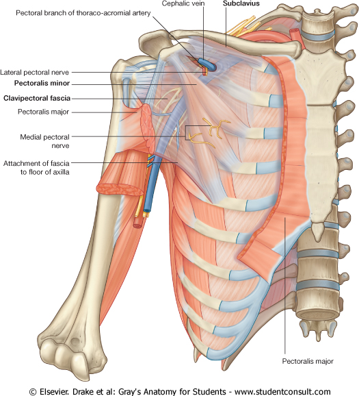
· Anterior wall of the axilla with the clavipectoral fascia (SlideShare)

· Section through the axilla: clavipectoral fascia and suspensory ligament (Blogger)
Related terms
- Infraclavicular region
- Clavicular region
- Deltopectoral triangle
- Pectoral fascia
- Pectoralis minor
- Subclavius
- Coracoid process
- Deltopectoral groove
- Cephalic vein
- Coracoclavicular ligament
References and sources
- Further reading: Clavipectoral fascia, Wikipedia.
- Federative International Programme for Anatomical Terminology (FIPAT). Terminologia Anatomica, 2nd ed. (TA2), 2019.