Proximal tubule (Latin: tubulus proximalis)
Overview
⇒ Structure of the whole kidney
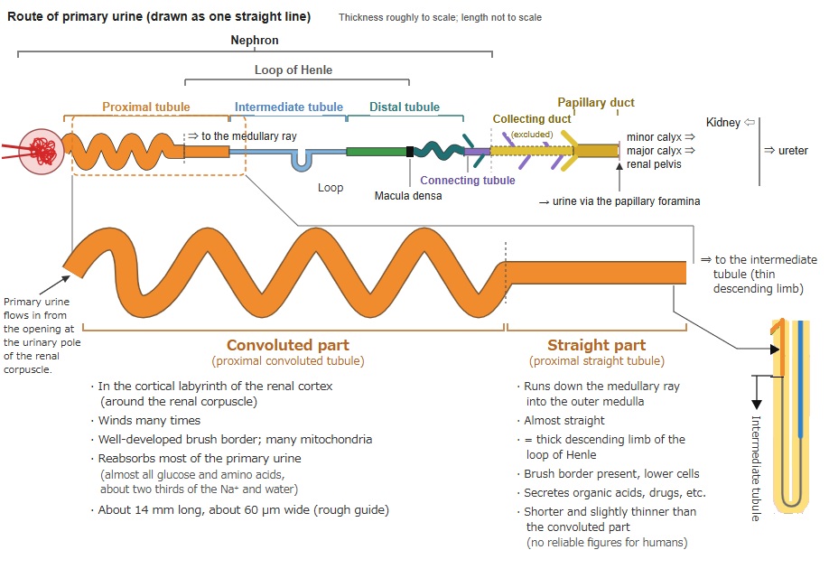
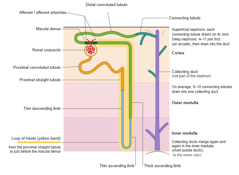
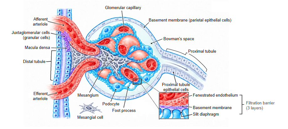
· The proximal tubule is the first part of the renal tubule. It starts at the urinary pole of the renal corpuscle and runs to the intermediate tubule (thin descending limb).
· In the nephron (renal corpuscle + renal tubule) it comes directly after the renal corpuscle.
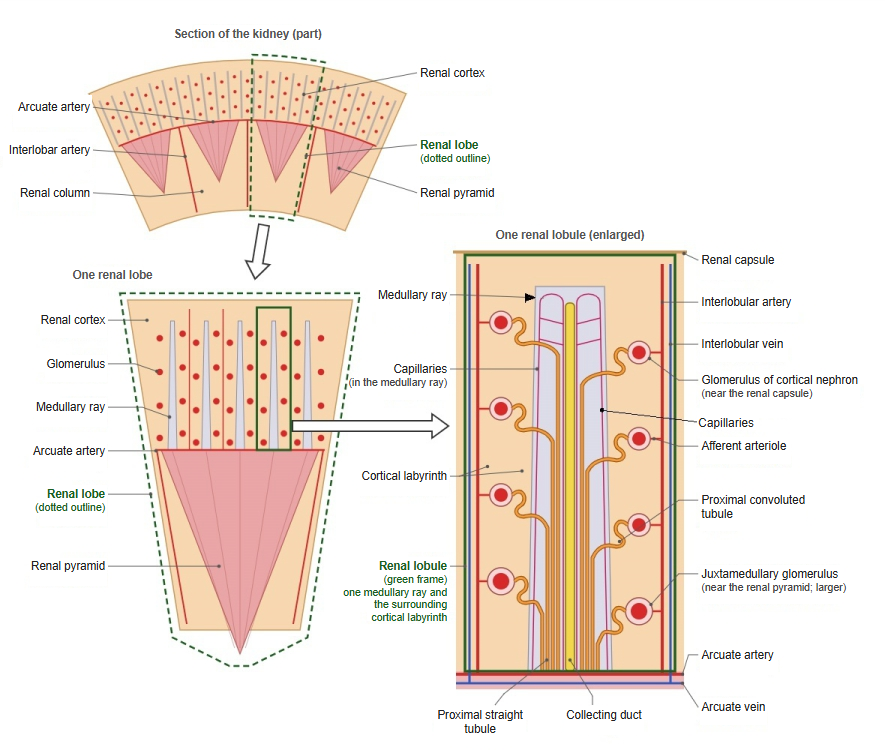
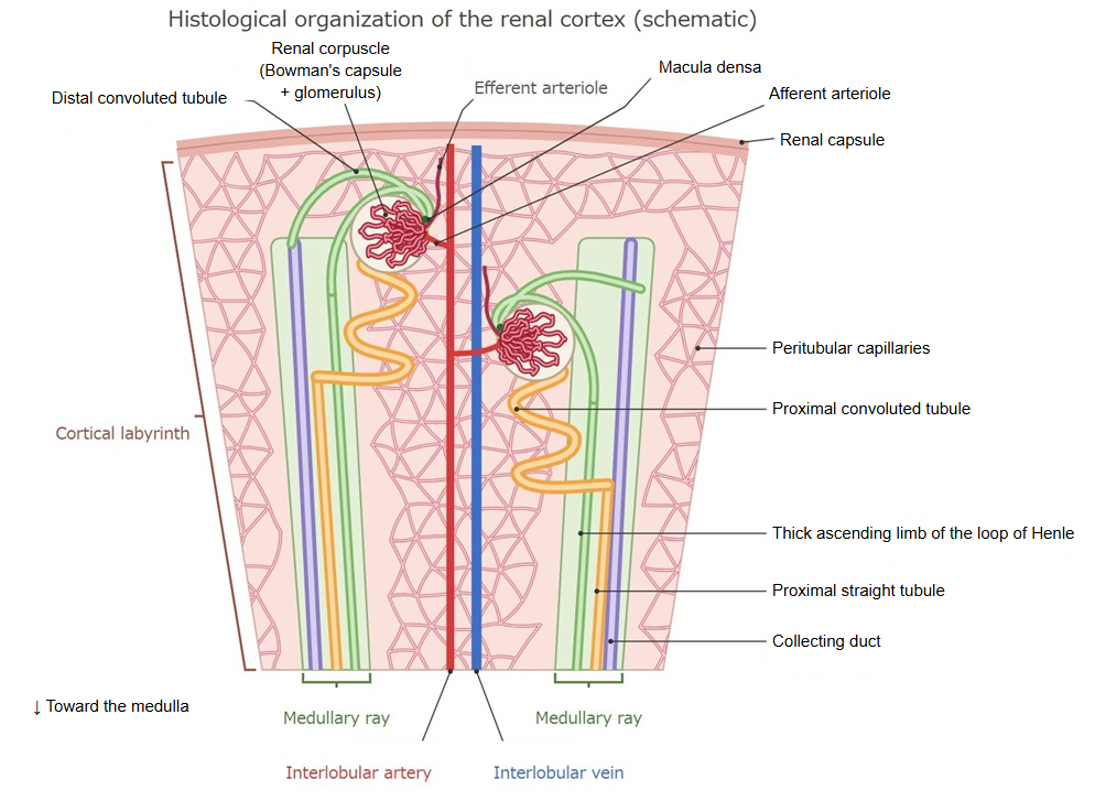
· It has a winding convoluted part (the proximal convoluted tubule) and a straight part that runs straight down (the proximal straight tubule, one of the straight tubules). The convoluted part lies in the cortical labyrinth; the straight part descends through a medullary ray into the outer zone of the renal medulla. The straight part is the first part of the loop of Henle.
· Its epithelium is simple cuboidal to low columnar, with strongly eosinophilic cytoplasm and a well-developed brush border of microvilli. In H&E sections it stains darker than the distal tubule.
· It reabsorbs most of the fluid filtered by the glomerulus (water, sodium, glucose and amino acids) back into the peritubular capillaries. By the secretion of hydrogen ions (acid) it also helps to regulate the pH of the primary urine.
· It is easily damaged by ischaemia and toxic drugs and is one of the main sites of acute tubular injury.
| Item | Convoluted part (proximal convoluted tubule) | Straight part (proximal straight tubule) |
|---|---|---|
| Position | Cortical labyrinth of the renal cortex (around the renal corpuscle) | Runs down in the medullary ray into the outer zone (outer stripe) of the medulla |
| Shape | Starts at the urinary pole of the renal corpuscle and winds many times | Runs almost straight down |
| Length / diameter | About 14 mm long (rough guide); diameter about 50–60 µm | Shorter than the convoluted part; no reliable figures |
| Epithelium | Tall simple cuboidal epithelium; well-developed brush border | Lower cells than in the convoluted part; brush border present |
| Cell base | Many basal infoldings and mitochondria | Fewer basal infoldings and mitochondria |
| Main work | Most of the reabsorption (almost all glucose and amino acids, about two thirds of the Na⁺ and water, HCO₃⁻) | Reabsorption (less than in the convoluted part); secretion of organic acids, drugs, etc. |
| Vulnerability | Relatively resistant to ischaemia (but easily damaged by drugs) | Easily damaged by lack of oxygen (ischaemia); the outer medulla is poor in oxygen |
| Continues into | The straight part | Intermediate tubule (thin descending limb) |
| Loop of Henle | Not included | Included (thick descending limb of the loop of Henle) |
· From differences in the cells, the proximal tubule is also divided into three segments, S1–S3 (convoluted part = S1 and S2; straight part = the end of S2 and S3). Items and figures differ between sources (some give about 14 mm as the length of the whole proximal tubule).
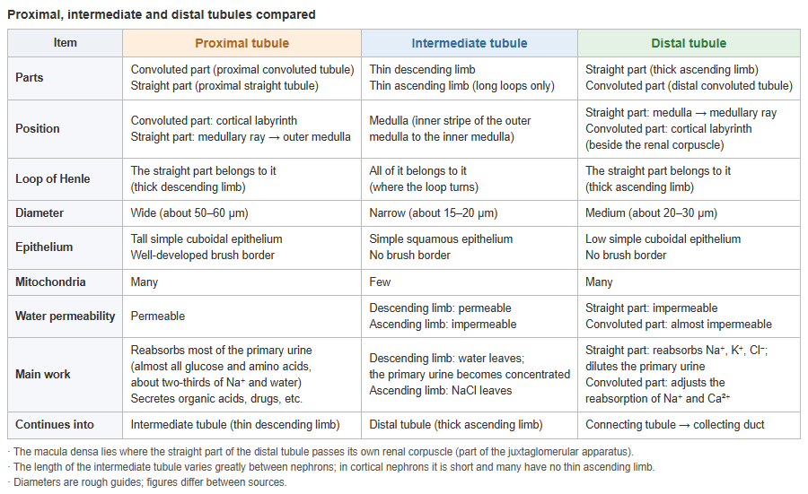
→ Proximal, intermediate and distal tubules compared (table)
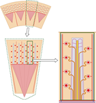 Renal lobe and renal lobule
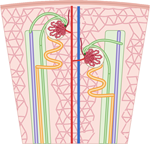 Tissue organization of the renal cortex
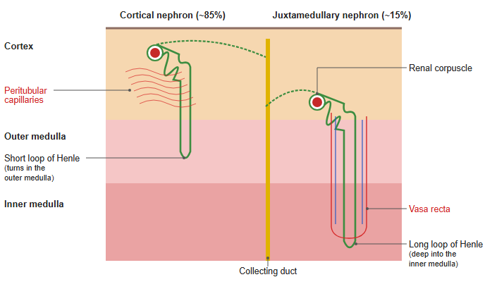
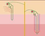 Cortical and juxtamedullary nephrons
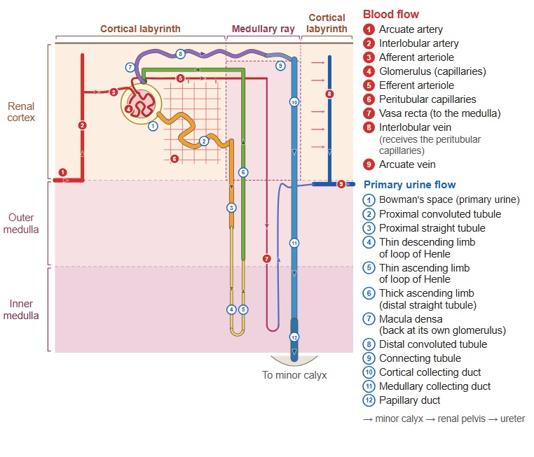
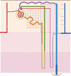 Nephron and blood vessels
Reference websites
[Wikipedia]
· Proximal tubule, Wikipedia.
· Nephron, Wikipedia.
[Websites with illustrations]
· Urinary system: kidney tubules (McGill Histology Atlas)
· Urinary system: renal cortex (McGill Histology Atlas)
· Histology of the tubules (Kidney Pathology)
· Kidney (virtual slide) (Histology Guide)
Related terms
- Straight tubule
- Distal tubule
- Cortical collecting duct
- Medullary collecting duct
- Papillary duct
- Kidney
- Renal parenchyma
- Renal artery
- Renal pelvis
- Urinary system
References and sources
- Further reading: Proximal tubule, Wikipedia.
- Further reading: Nephron, Wikipedia.
- Federative International Programme for Anatomical Terminology (FIPAT). Terminologia Anatomica, 2nd ed. (TA2), 2019.