Glomerular basement membrane
Overview
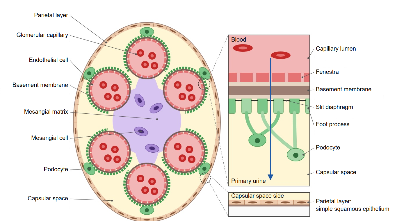
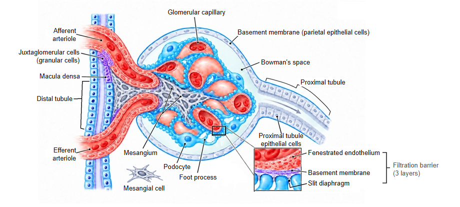
· The glomerular basement membrane is the thick basement membrane between the endothelial cells of the glomerular capillaries and the podocytes. It is the central layer of the filtration barrier of the glomerulus.
· It forms by fusion of the basal laminae of the capillary endothelium and of the podocytes, so it is thicker than basement membranes elsewhere (about 250-400 nm; sources differ). On its outer side lie the podocytes that make up the visceral layer of the glomerular capsule, which encloses the glomerulus within the renal corpuscle.
· Under the electron microscope it shows three layers, from the endothelial side: the lamina rara interna, the lamina densa and the lamina rara externa.
· It contains mainly type IV collagen, laminin and heparan sulfate. It is made and maintained by the endothelial cells and the podocytes.
· The primary urine (filtrate) passes in turn through the pores of the endothelium, this basement membrane and the gaps between the podocyte foot processes (the filtration slits) into the capsular space. The membrane sieves by size and charge and holds back albumin and other plasma proteins.
· When the membrane or the filtration barrier is damaged, protein leaks into the urine (proteinuria). The membrane becomes thickened in membranous nephropathy, and in Alport syndrome a defect in a type IV collagen gene makes it fragile.
Reference websites
[Wikipedia]
· Glomerular basement membrane, Wikipedia.
· Glomerulus (kidney), Wikipedia.
[Websites with illustrations]
· Filtration barrier (diagram) (Wikimedia Commons)

· Renal corpuscle (electron micrograph) (Histology Guide)
· The glomerular basement membrane (review article) (PubMed Central)
· Renal corpuscle (diagram) (Wikimedia Commons)

Related terms
- Glomerular capillary
- Podocyte
- Visceral layer of glomerular capsule
- Parietal layer of glomerular capsule
- Capsular space
- Mesangium
- Vascular pole
- Urinary pole
- Kidney
- Renal parenchyma
- Interlobular arteries of kidney (cortical radiate arteries)
- Renal artery
- Urinary system
References and sources
- Further reading: Glomerular basement membrane, Wikipedia.
- Further reading: Glomerulus (kidney), Wikipedia.
- Federative International Programme for Anatomical Terminology (FIPAT). Terminologia Anatomica, 2nd ed. (TA2), 2019.