Antitragohelicine fissure (Latin: fissura antitragohelicina)
The fissure cuts the cauda helicis, the lower tail-like end of the helix cartilage, off from the cartilage of the antitragus and the antihelix. It lies above the lobule, which has no cartilage. In the living ear it is filled by fibrous tissue and covered by skin. It is seen most clearly on the medial (cranial) surface of the isolated cartilage.
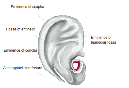
Names of the parts of the auricle
The surface of the auricle is full of ridges and hollows, and its parts have the following names.
| Lateral surface | 1 | Helix | 8 | Superior crus of antihelix | 15 | Tragus | Medial surface | 1 | Eminence of scapha |
| 2 | Auricular tubercle | 9 | Triangular fossa | 16 | Anterior notch | 2 | Eminence of concha | ||
| 3 | Crus of helix | 10 | Concha of auricle | 17 | External acoustic meatus | 3 | Fossa of antihelix | ||
| 4 | Spine of helix | 11 | Cymba of concha | 18 | Scaphoid fossa | 4 | Eminence of triangular fossa | ||
| 5 | Lobule of auricle | 12 | Cavity of concha | 19 | Tail of helix | 5 | |||
| 6 | Antihelix | 13 | Antitragus | 20 | Antitragohelicine fissure | ||||
| 7 | Inferior crus of antihelix | 14 | Intertragic notch |
Note: the antitragohelicine fissure can also be seen from the lateral surface.
Reference websites
[Wikipedia]
· Auricle (anatomy), Wikipedia.
[Websites with illustrations]
· Outer surface of the cartilage of the auricle (vintage engraving) (Dreamstime)

· Cartilage of the auricle (Clinical Gate)
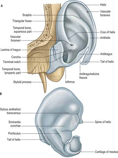
· Auricle and its cartilage (Pocket Dentistry)
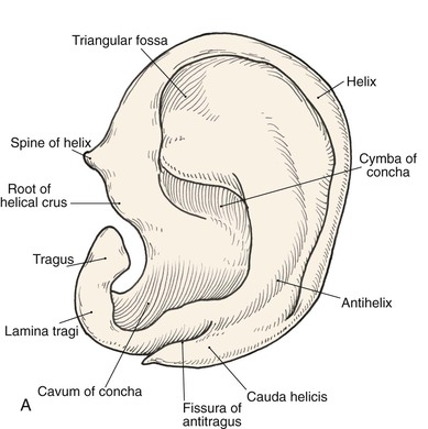
· The auricle, lateral surface (Gray's Anatomy, fig. 904) (Wikimedia Commons)

Related terms
References and sources
- Further reading: Auricle (anatomy), Wikipedia.
- Federative International Programme for Anatomical Terminology (FIPAT). Terminologia Anatomica, 2nd ed. (TA2), 2019.