Pulmonary alveoli (Latin: alveoli pulmonis)
Also called the air sacs (singular: pulmonary alveolus)
Overview
· The alveoli are the centre of the work of the respiratory system and the most important part of the lung parenchyma (the functional tissue of the lung).
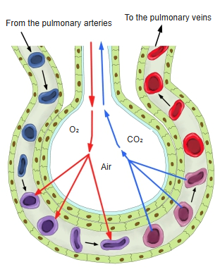
· Neighbouring alveoli lie very close together: two adjacent alveoli share, from both sides, the single capillary network inside the alveolar septum between them.
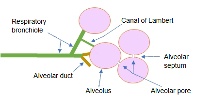
- Conducting part : main bronchus ~ lobar bronchus ~ segmental bronchus ~ terminal bronchiole
- Respiratory part : alveolar ducts, alveoli, alveolar sacs
From the main bronchus to the alveoli
The segmental bronchi also give off segmental bronchial branches.
Blood flow through the lung
The pulmonary arteries carry venous blood and the pulmonary veins carry arterial blood.
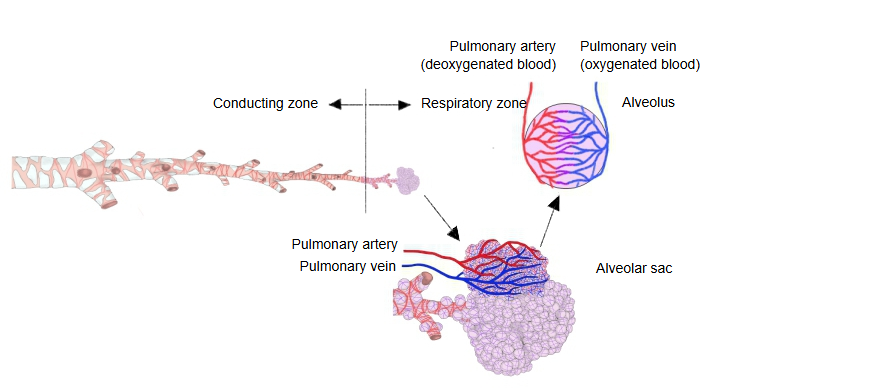
· The surface area of one alveolus is about 270,000 µm² (0.27 mm²); all the alveoli of one person together give a surface of about 70–80 m².
Another common description of the alveolus:
An alveolus is formed by very thin, squamous respiratory epithelial cells (the alveolar cells), about 0.1 µm thick,
which form a small ball about 0.1–0.2 mm across around a cavity filled with air (the alveolar air).
· English Wikipedia gives a larger diameter for an alveolus: 200–500 µm (sources differ).
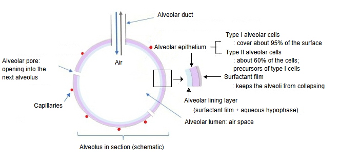
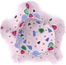 Alveolus (section)
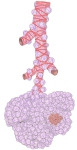 Alveolar sac
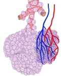 Alveoli and blood vessels
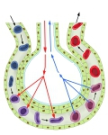 Gas exchange
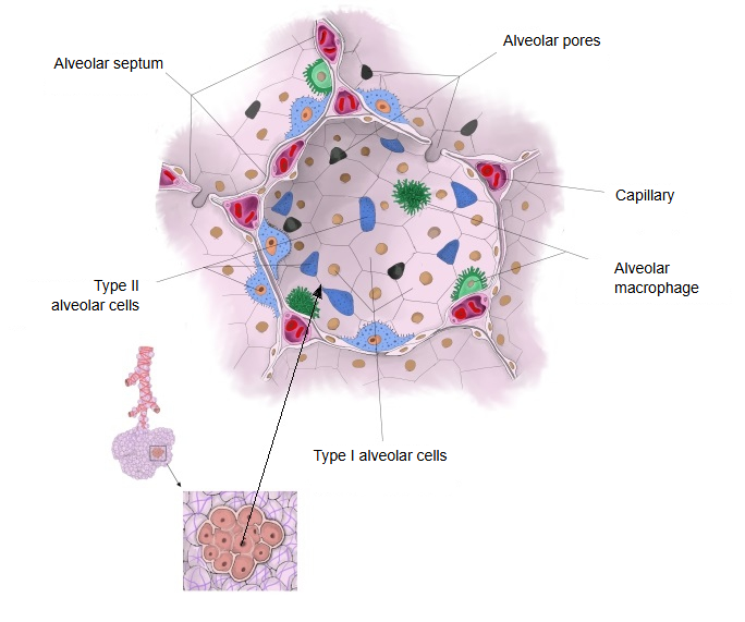
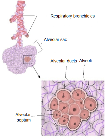
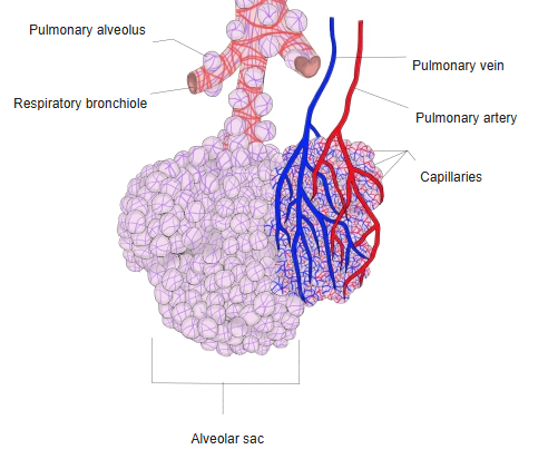
Structure
of the
alveolus
- Alveolar cavity : the space that holds the air.
- What forms the alveolar cavity
- Alveolar lining layer : the innermost layer; a protective layer that keeps the epithelial cells from touching the air directly.
- Alveolar epithelial cells : type I (the main site of gas exchange) and type II cells; together they make up 99% of the epithelial cells.
- Surface film : a single layer of phospholipids (including unsaturated fatty acids); keeps the shape of the alveolus.
The features of the three types of alveolar epithelial cells are listed below.
Besides these three types, the alveoli also contain phagocytes called alveolar macrophages (or dust cells), which wander over the surface and take up particles; they are not part of the epithelium.
| 1 | Type I alveolar cell | · Also called squamous alveolar epithelial cells. · Share: 39% of the cells, but each cell covers about 36 times the area of a type II cell, so together they cover about 96% (95.8%) of the alveolar surface. · Very thin, flat cells: the cytoplasm spreads about 100 µm wide; thickness about 0.05–0.2 µm (sources differ). · Neighbouring cells are joined by tight junctions (zonulae occludentes). |
| 2 | Type II alveolar cell | · Share: 60% of the cells. · Also called granular pneumocytes or great alveolar cells. · Cuboidal cells with short microvilli at the apex. · Secrete surfactant ⇒ the main component of the lining layer. · Can divide and differentiate into type I cells. |
| 3 | Type III alveolar cell | · Share: less than 1%. · Cuboidal (brush) cells with thick microvilli at the apex. · Usually found beside type II cells. |
· The alveolar lining layer lies on the inner side of the alveolar epithelial cells.
· Because of this layer, the alveolar epithelial cells do not touch the air of the alveolar cavity directly.
· Its thickness varies from place to place, from about 50 nm to a few micrometres.
· It is made mainly by type II alveolar cells, with some contribution from type I cells.
· It has two layers: the surface film (the innermost layer, facing the air) and a watery base layer next to the cells.
Surface film
· A single layer of phospholipids (surfactant), including some with unsaturated fatty acids.
· It keeps the shape of the alveoli: by lowering the surface tension it stops them from collapsing.
Reference websites
[Wikipedia]
· Pulmonary alveolus, Wikipedia.
· Pulmonary surfactant, Wikipedia.
[Websites with illustrations]
· Alveoli (illustration) (Verywell Health)

· Alveolus diagram (Wikimedia Commons)

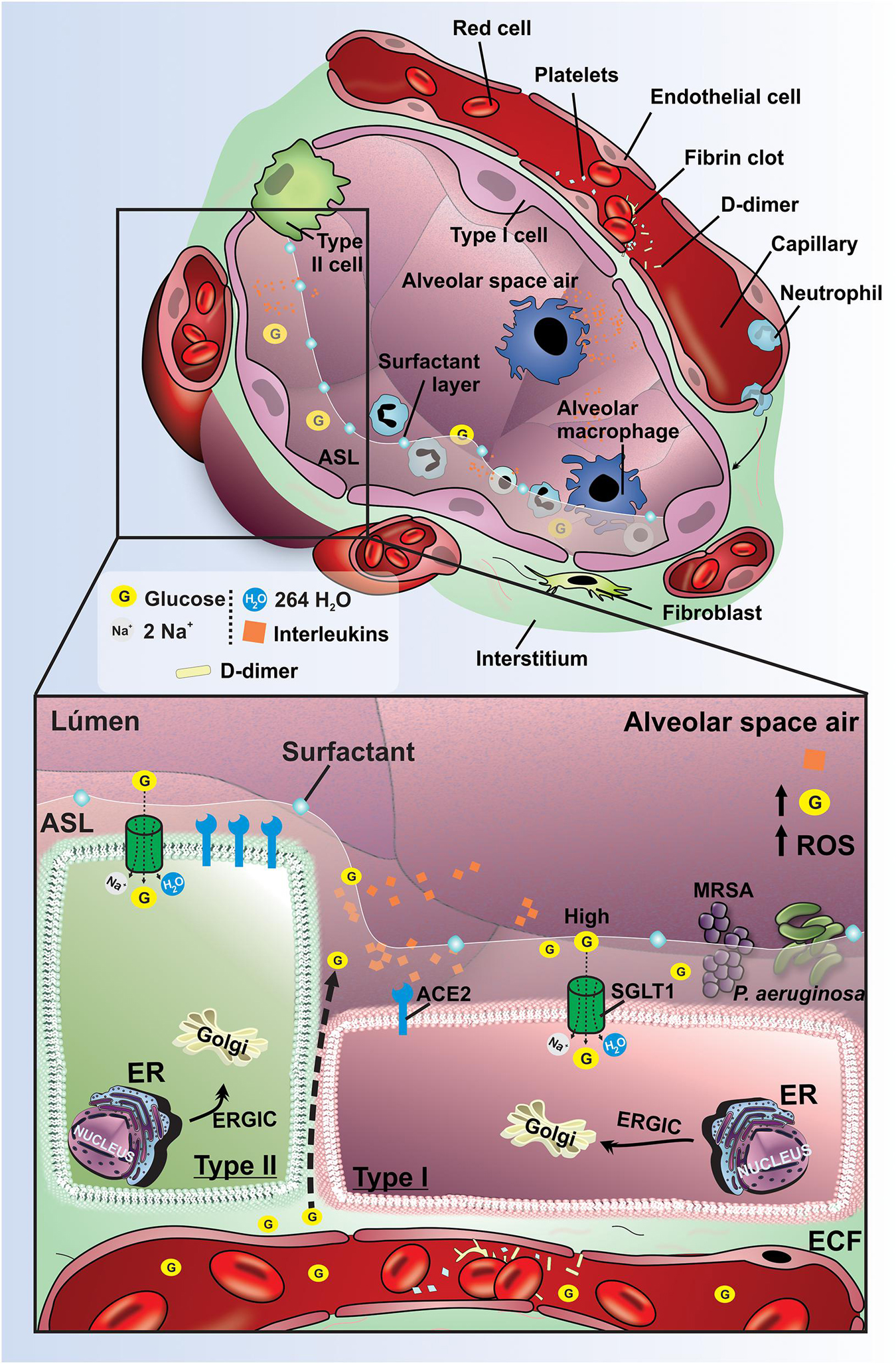
· Structure of the alveolus (Frontiers in Physiology)
Related terms
- Alveolar septum
- Blood
- Alveolar sac
- Alveolar duct
- Alveolar pores
- Alveolar cells
- Alveolar lining layer
- Respiratory bronchiole
- Lung
References and sources
- Further reading: Pulmonary alveolus, Wikipedia.
- Further reading: Pulmonary surfactant, Wikipedia.
- Federative International Programme for Anatomical Terminology (FIPAT). Terminologia Anatomica, 2nd ed. (TA2), 2019.