Retinal pigment epithelium (Latin: stratum pigmentosum retinae)
Also called RPE or the pigmented layer of retina or the pigment epithelium
Note: in this site's eyeball pages the inner layer of the eyeball is divided into the retina and the pigment epithelium layer. Many sources do not separate them and count the pigment epithelium as the outermost layer of the retina. It develops from the outer wall of the optic cup, while the neural retina develops from the inner wall.
- Retina : made up of the optic part and the blind part; only the optic part senses light, and it has many layers.
- Pigment epithelium layer : three parts: the retinal pigment epithelium, the pigment epithelium of the ciliary body and the pigment epithelium of the iris; each is a single layer of pigmented epithelium.
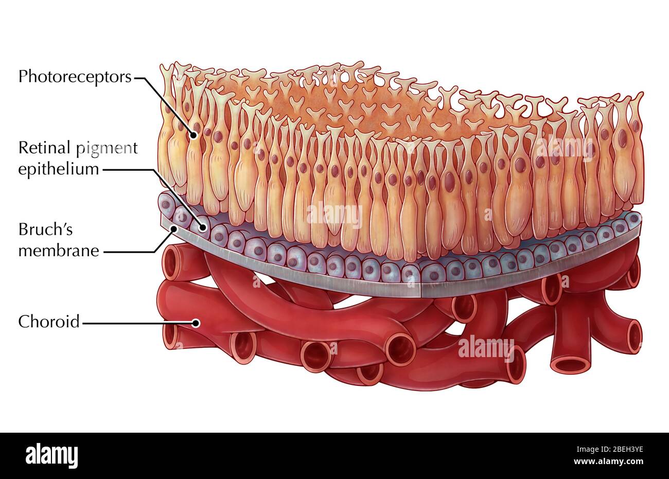
- Retinal pigment epithelium : hexagonal prism-shaped cells, firmly joined to each other; send microvilli-like processes towards the photoreceptor cells.
- Ciliary pigment epithelium : covers the inner surface of the ciliary body; cuboidal cells; thinner than the retinal pigment epithelium, with no microvilli-like processes.
- Iridial pigment epithelium : short columnar cells covering the back of the iris; at the pupillary margin it passes into the iridial part of the retina.
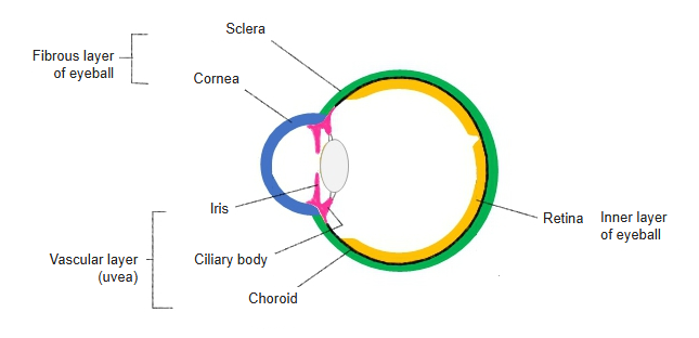
Pigment epithelium layer (inner layer of the eyeball), from back to front
Other functions of the retinal pigment epithelium include:
· absorbing stray light that enters the retina, by its pigment granules, which reduces scatter inside the eye;
· epithelial transport: carrying nutrients from the choroidal capillaries to the photoreceptors and removing water, ions and waste from the space around them.
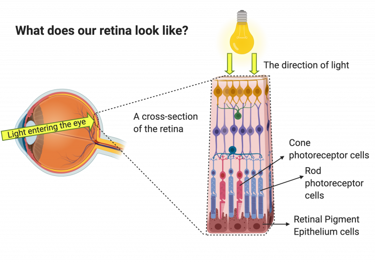
It also recycles the visual pigment (retinal) used by the rods and cones (the visual cycle), engulfs and digests the tips of the photoreceptor outer segments that are shed every day, and releases growth factors that help keep the eye immune-privileged. See also the Wikipedia article.
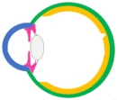 Layers of the eyeball wall
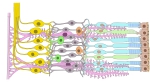 Layers of the retina
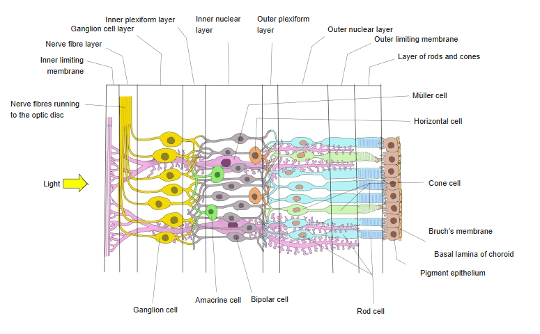
Over the retina the pigment cells are hexagonal prisms. Well-developed junctions between neighbouring cells keep the tissue fluid of the choroid out of the retina and limit the spread of inflammation into it. The base of each cell, which rests on the basal lamina of the choroid and holds the nucleus, has no pigment granules; the part above the nucleus is filled with melanin granules and sends microvilli-like processes in between the photoreceptor cells of the retina. The attachment between the pigment epithelium and the photoreceptors is weak; this is the plane that separates in retinal detachment.
Reference websites
[Wikipedia]
· Retinal pigment epithelium, Wikipedia.
[Websites with illustrations]


Related terms
- Choroidocapillary lamina
- Retina
- Optic part of retina
- Ciliary part of retina
- Iridial part of retina
- Outer segment of photoreceptor
- Basal lamina of choroid
- Choroid
References and sources
- Further reading: Retinal pigment epithelium, Wikipedia.
- Further reading: Retina, Wikipedia.
- Federative International Programme for Anatomical Terminology (FIPAT). Terminologia Anatomica, 2nd ed. (TA2), 2019.