Cartilage (Latin: cartilago)
Overview
Cartilage is one of the tissues that build the body; in evolution it is thought to be the tissue that served as the model for bone.
Tissues: cartilage is a supporting tissue, one of the connective tissues: firm but flexibleThe model for bone: most bones of the skeleton are still first formed as cartilage models that are later replaced by bone; the epiphyseal cartilage (growth plate) is the part that remains during growth
Cartilage is formed by the cartilage cells (chondrocytes) it contains (only about 1–5% of its volume), and is classified into the following three types.
Chondrocytes: the cells secrete fibres and glycoproteins that form the framework (matrix) of cartilage
Types
of
cartilage
of
cartilage
- Hyaline cartilage : the most common type; a glassy, translucent, uniform matrix with moderate hardness and elasticity. Cells 3–5%, mainly fine type II collagen fibres, water about 70–80%. Sites: articular cartilage, costal cartilage, tracheal and bronchial cartilages, nasal cartilages, most laryngeal cartilages.
- Elastic cartilage : yellowish, flexible and highly elastic; it bends and springs back. Cells 2–5%, a dense network of elastic fibres (elastin) plus type II collagen fibres, water about 60–70%. Sites: auricle, external acoustic meatus and auditory tube, epiglottis.
- Fibrocartilage : whitish and the toughest of the three; resists compression and tension. Cells 1–2%, thick bundles of type I collagen fibres, water about 50–65%; no perichondrium. Sites: intervertebral discs, pubic symphysis, menisci, acetabular labrum and other articular labra.
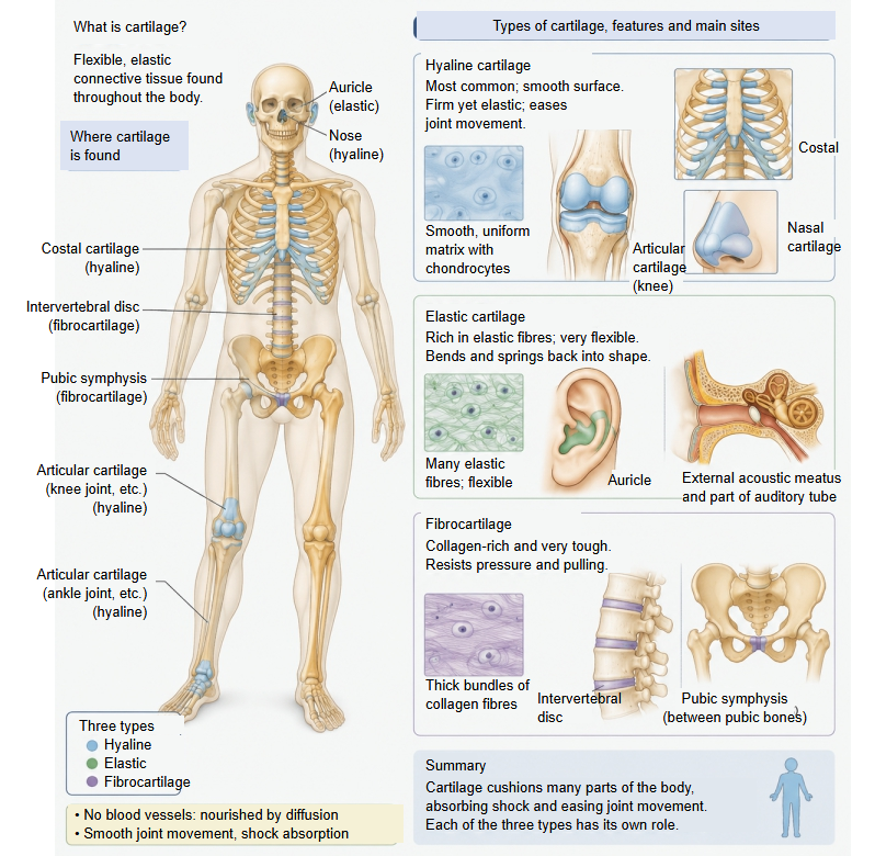
Cartilage is a special tissue in which no blood vessels, nerves or lymphatic vessels are found at all.
Most tissues receive oxygen and nutrients from blood vessels that enter them, but no blood vessels enter cartilage.
Oxygen and nutrients therefore reach its cells by diffusion through the surrounding fluid: synovial fluid, or tissue fluid from the vessels of the perichondrium. For this reason cartilage repairs poorly.
Its matrix resists compression and gives elasticity, so cartilage supports soft structures (nose, airways, ear), cushions joints and joins bones in cartilaginous joints.
Composition
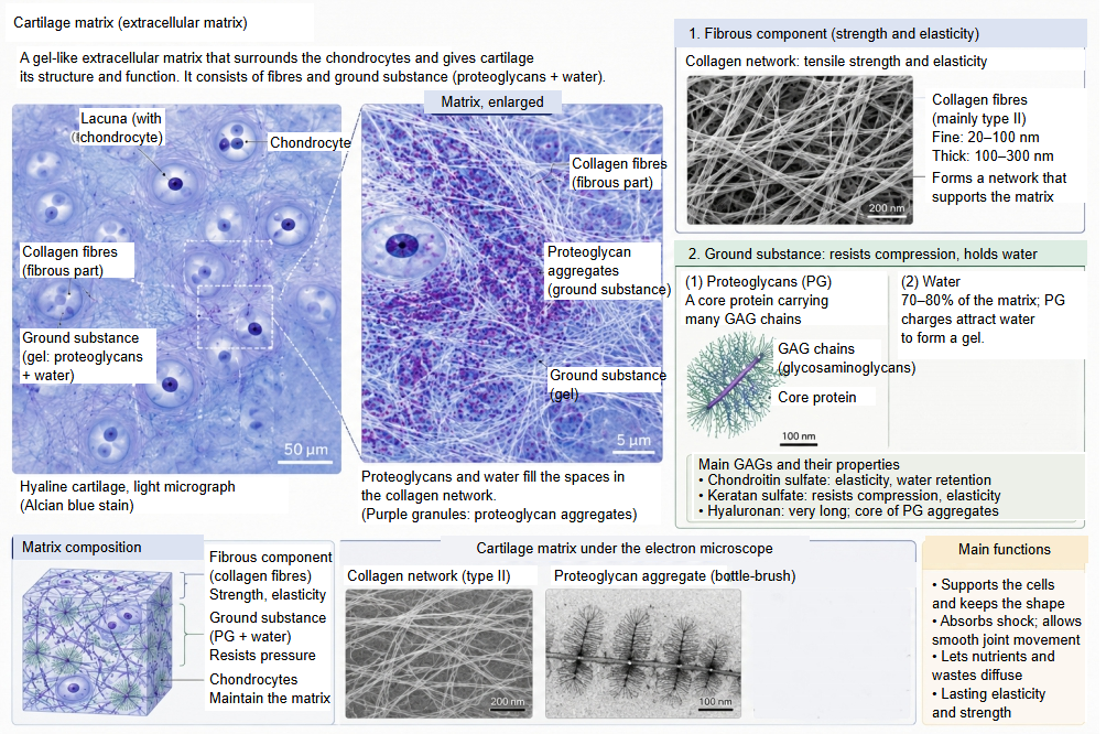
Cartilage consists of a cellular component and everything else, the cartilage matrix; apart from the water, the matrix is all produced by the cartilage cells.
Cellular component: the chondrocytes, which develop from mesenchymal precursor cells
Composition
of
cartilage
of
cartilage
- Cartilage cells : chondrocytes: produce the cartilage matrix (everything except the water) and maintain the function of cartilage; about 1–5% of the volume. Each lies in a small space, the lacuna.
- Cartilage matrix : = extracellular matrix
- Fibrous component : mainly type II collagen (hyaline cartilage); also type I collagen (fibrocartilage) and elastic fibres (elastic cartilage).
- Ground substance : amorphous: proteoglycans (most of it, chiefly aggrecan), hyaluronan and glycoproteins.
- Water : about 60–80% of the matrix, held by the proteoglycans; important for the diffusion of nutrients and wastes.
Related terms
- Symphysis
- Collagen fiber
- Cartilage matrix
- Cartilage lacuna
- Perichondrium
- Articular cartilage
- Costal cartilage
- Epiphyseal cartilage
- Cartilaginous joint
- Intervertebral disc
- Auricular cartilage
References and sources
- Further reading: Cartilage, Wikipedia.
- Further reading: Chondrocyte, Wikipedia.
- Federative International Programme for Anatomical Terminology (FIPAT). Terminologia Anatomica, 2nd ed. (TA2), 2019.