Hyaline cartilage (Latin: cartilago hyalina)
- Overview
- Composition
- Cell arrangement
- Where it is found
- Comparison of the three types of cartilage
- Related terms
Overview
Hyaline cartilage is one of the three types of cartilage, and the one most common in the body.
Cartilage: a special tissue with no nerves, blood vessels or lymphatics; its cells are nourished by diffusion through the water-rich matrix
In the living body it is translucent and milky white; because it holds much water, it absorbs shock well and gives a smooth, low-friction surface.
Milky white: or faintly bluish; the name comes from the Greek word for glass (hyalos)
Types
of
cartilage
of
cartilage
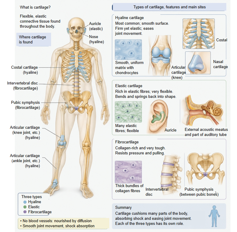
- Hyaline cartilage : most cartilage is of this type: a translucent, uniform matrix, fairly hard and elastic. Make-up: cells about 1–5%, mainly type II collagen fibres, water about 70–80%. Sites: articular, costal, tracheal and bronchial, nasal cartilages.
- Elastic cartilage : yellowish, flexible and elastic; springs back after bending. Make-up: cells about 2–5%, elastic fibres (elastin) + type II collagen fibres, water about 60–70%. Sites: auricle, external acoustic meatus, auditory tube, epiglottis.
- Fibrocartilage : whitish, the toughest of the three; resists pressure and pulling. Make-up: cells about 1–2%, thick bundles of type I collagen fibres, water about 50–65%. Sites: intervertebral discs, pubic symphysis, menisci, labra.
Composition
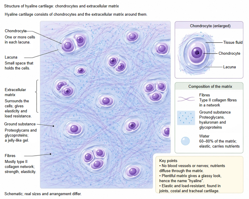
Hyaline cartilage contains about 70–80% water, while its cells make up only about 1–5%.
Water: the most of any type of cartilage, held by the proteoglycans
The table below briefly sums up what hyaline cartilage is made of.
| Component | Share (approx.) | Role and features | ||
| Chondrocytes | 1–5% | The only cells of cartilage; each lies in a small space, the lacuna. They secrete the extracellular matrix (everything below except the water) and keep the cartilage working. | ||
| Extracellular matrix | Water | 70–80% | The largest component of the cartilage: it absorbs shock under load, makes the cartilage spring back, and carries oxygen and nutrients to the cells. | |
| Collagen (= collagen fibres) | 10–15% | Mainly type II collagen (over about 90% of it). It forms a fine network that gives tensile strength and is the framework that traps the water-holding molecules. The fibres are so fine that the matrix looks uniform under the light microscope. | ||
| Proteoglycans | 5–10% | Giant molecules (mainly aggrecan) in which many sugar chains (chondroitin sulfate and keratan sulfate) are bound to a core protein; they carry a strong negative charge and attract and hold water strongly. | ||
| Other glycoproteins | 1–3% | Link protein, fibronectin, anchorin CII and others; they bind and anchor collagen, proteoglycans and cells to each other. | ||
Cell arrangement
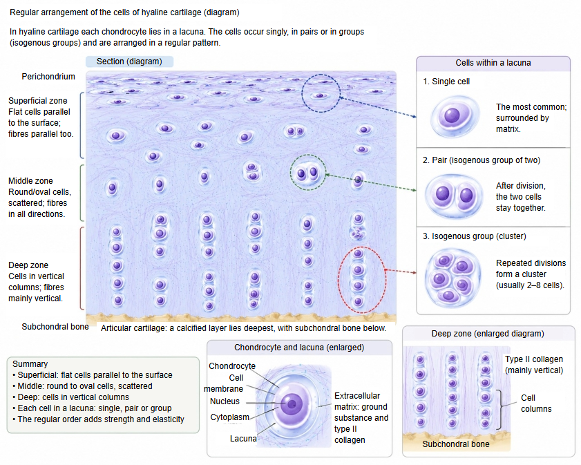
The arrangement of the chondrocytes is typical for each type of cartilage, and it also differs between sites in the same type. Chondrocytes occur singly, in pairs or in small clusters (isogenous groups) formed by repeated division of one cell.
In articular cartilage the chondrocytes are arranged regularly, from flattened cells parallel to the surface in the superficial layer to vertical columns in the deep layer.
Middle zone: rounded cells scattered between them; below the deep zone lies a thin layer of calcified cartilage, resting on the subchondral bone
Where it is found
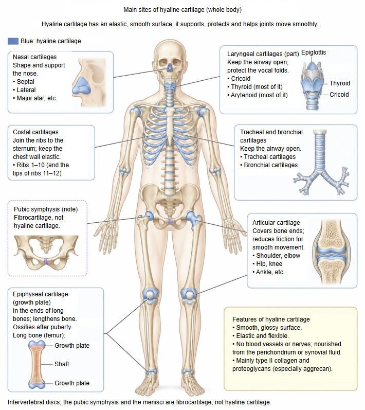
Nasal cartilages, costal cartilages, tracheal (and bronchial) cartilages, articular cartilage, epiphyseal cartilage and part of the laryngeal cartilages are hyaline cartilage.
| Site | Examples | Main role and features | |
| 1 | Articular cartilage | Joint surfaces of the synovial joints of the limbs, the vertebral column and elsewhere | Reduces friction and absorbs shock. It has no perichondrium. |
| 2 | Costal cartilages | Front ends of the 1st–10th ribs | Give the thoracic cage elasticity and help the breathing movements. |
| 3 | Nasal cartilages | Septal cartilage, lateral nasal cartilage, major alar cartilage and others | Keep the shape of the nose. |
| 4 | Laryngeal cartilages | Cricoid cartilage, most of the thyroid cartilage, most of the arytenoid cartilages | Keep the airway open and take part in voice production. They tend to calcify and ossify with age. |
| 5 | Tracheal and bronchial cartilages | Tracheal cartilages, and the cartilages of the main bronchi, lobar bronchi and smaller bronchi | Keep the airway open and make breathing easy. |
| 6 | Epiphyseal cartilage (growth plate) | Ends (epiphyses) of growing long bones | Drives the growth of the bone in length; replaced by bone in adults. Most bones are first laid down as hyaline cartilage models. |
Comparison of the three types of cartilage
Below is a simple table comparing the three types of cartilage.
| Item | Hyaline cartilage | Elastic cartilage | Fibrocartilage | |
| 1 | Appearance | Translucent and smooth; moderately hard and elastic. | Yellowish and very elastic; springs back strongly. | Whitish and very tough; resists both pressure and pulling. |
| 2 | Cells | About 1–5% (sources differ) | About 2–5% (some sources give 10%) | About 1–2% |
| 3 | Water | The most (about 70–80%) | About 60–70% | The least (about 50–65%) |
| 4 | Fibres | Type II collagen fibres | Elastic fibres (elastin) + type II collagen fibres | Type I collagen fibres in thick bundles + some type II |
| 5 | Perichondrium | Present (except on articular cartilage) | Present | Absent |
| 6 | Function | Support, shock absorption, smooth joint movement | Keeps flexibility and shape | Resists strong pressure and pulling |
| 7 | Sites | Articular cartilage, costal cartilages, nasal cartilages, tracheal and bronchial cartilages, epiphyseal cartilage, part of the laryngeal cartilages (cricoid, thyroid, arytenoid) | Auricular cartilage, external acoustic meatus, auditory tube, part of the laryngeal cartilages (epiglottic, corniculate, cuneiform, sesamoid) | Intervertebral discs, pubic symphysis, menisci, articular discs, labra, and the attachments of some tendons and ligaments |
Related terms
- Synchondrosis
- Cartilage
- Cartilage matrix
- Cartilage lacuna
- Perichondrium
- Articular cartilage
- Costal cartilage
- Epiphyseal cartilage
References and sources
- Further reading: Hyaline cartilage, Wikipedia.
- Further reading: Cartilage, Wikipedia.
- Federative International Programme for Anatomical Terminology (FIPAT). Terminologia Anatomica, 2nd ed. (TA2), 2019.