Cell membrane (Latin: Plasmalemma)
Also called the plasma membrane
- Overview
- Structure (Phospholipid bilayer, Membrane proteins, transmembrane proteins, peripheral proteins, lipid-anchored proteins, Sugar chains, bound to proteins, bound to lipids, Cholesterol)
- Fluidity
- Q: Are the phospholipids of the membrane constantly renewed?
- Reference websites
- (Wikipedia, websites with illustrations)
Overview
of the
cell
- Cell membrane : phospholipid bilayer, membrane proteins, sugar chains
- Cytoplasm
- Cytosol : water, electrolytes, metabolic enzymes, free amino acids, metabolic intermediates
- Organelles : mitochondria, endoplasmic reticulum, centrosome, Golgi apparatus, lysosomes, peroxisomes, vesicles (some sources do not count vesicles as organelles)
- Cytoskeleton : microtubules, actin filaments, intermediate filaments
- Nucleus : nuclear envelope, nucleoplasm, chromatin, nucleolus; the nucleus is also counted among the organelles
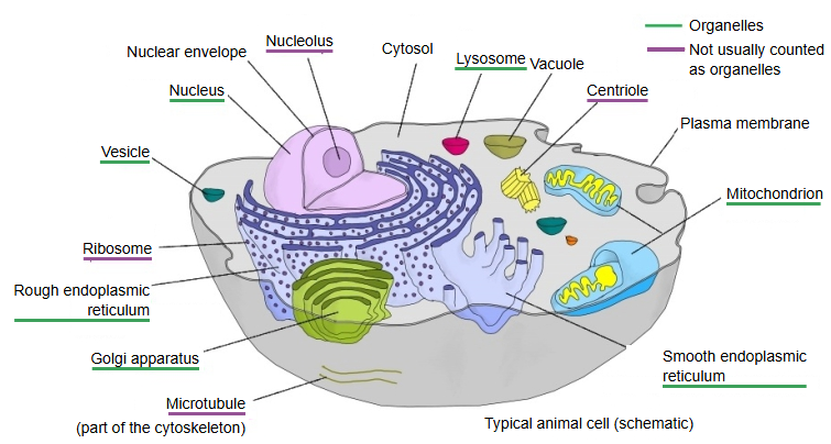
Structure
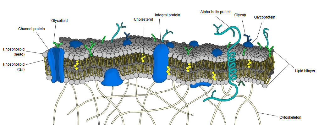
The structure of the cell membrane can be divided broadly into the following four parts.
structure
of the cell
membrane
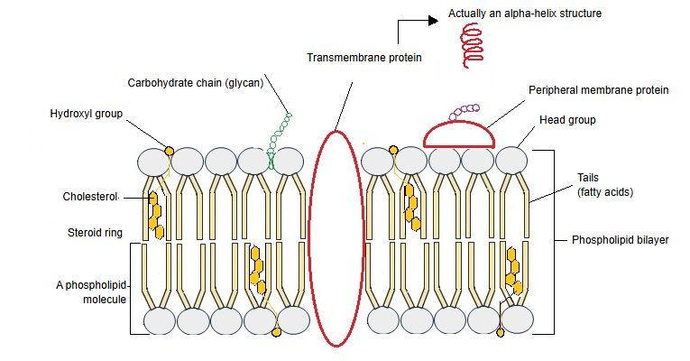
- Phospholipid bilayer : the hydrophilic heads face outwards, the hydrophobic tails inwards.
- Membrane proteins : receptors, channels, transporters, enzymes and others.
- Sugar chains : (glycolipids, glycoproteins) take part in cell recognition and adhesion.
- Cholesterol : regulates the fluidity of the membrane.
The cell surface has a complex structure, and its features differ from one kind of cell to another.
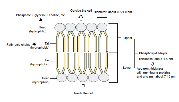
Phospholipid bilayer
lipid
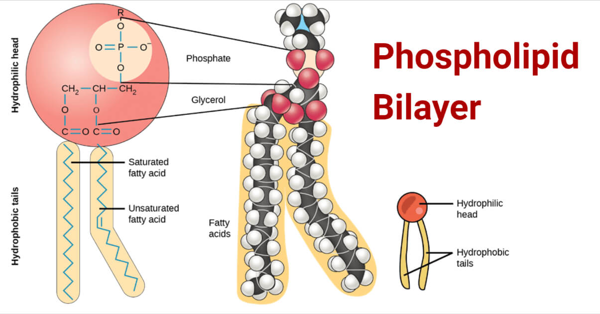
- Head : almost spherical, about 0.8–1.0 nm across. Make-up: phosphate group, glycerol backbone, polar group.
- Tails : two fatty-acid chains; usually one is a saturated and the other an unsaturated fatty acid.
Bilayer: the phospholipids of the upper and lower layers are arranged with their tails facing each other, forming two layers.
Functions:
· It forms the basic boundary of the cell.
· It is selectively permeable and blocks water-soluble substances. It is fluid, which allows membrane proteins to work and vesicles to be transported.
About fluidity:
Renewal of the membrane molecules: old phospholipid molecules are replaced by new ones made mainly in the endoplasmic reticulum; the replaced molecules are often reused in the cell.
Fluidity means that the membrane is not a solid plate: its molecules can move within its plane, as in a liquid. As a result:
· membrane proteins can work;
· receptors and transporters can gather together or spread apart;
· the membrane can be repaired, fuse with other membranes and divide.
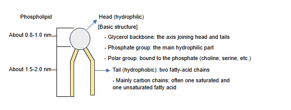
What the hydrophilic head means:
· A website with an illustration showing the detailed structure of the heads and tails (Microbe Notes)
Membrane proteins
proteins
- Transmembrane proteins : a very common type; they pass through the membrane and act as channels, transporters and receptors. In the plasma membrane their membrane-spanning parts are almost always alpha-helices.
- Membrane surface proteins
- Peripheral proteins : bound to the membrane surface temporarily.
- Lipid-anchored proteins : the protein itself lies outside the lipid layer and is fixed to the membrane by a lipid that acts as an anchor.
- Note : alpha-helix (spiral) structure: many illustrations draw channel proteins without a spiral, but this is a simplified drawing; the membrane-spanning parts of the transmembrane proteins of the human plasma membrane are alpha-helices. (Beta-barrel membrane proteins occur in the outer membranes of mitochondria.)
⟨ Functions of the transmembrane proteins ⟩
· Transport of substances (ion channels, glucose transporters and others).
· Receiving signals (G-protein-coupled receptors (GPCRs), receptor tyrosine kinases).
· Adhesion between cells (cadherins, integrins).
Features: they connect the environments outside and inside the cell, and are structurally anchored firmly in the membrane.
⟨ Functions of the peripheral proteins ⟩
· Relaying signals (e.g. kinases, G proteins).
· Linking to the cytoskeleton (e.g. actin-binding proteins).
· Controlling the activity of enzymes.
Features: they can come off the membrane and are very mobile; they are suited to regulation and signalling.
⟨ Functions of the lipid-anchored proteins ⟩
· Signal transmission (e.g. Ras, Src).
· Local gathering and fixing on the membrane.
· Keeping their function as receptors or enzymes.
Features: they stay stably on the membrane and often work in particular regions of it.
Sugar chains
By far most of the sugar chains are covalently bound to proteins (glycoproteins).
⟨ Bound to proteins ⟩
1. Definition: sugar chains covalently bound to a protein.
Position: as a rule they project from the outer side of the cell.
Examples: N-linked: the sugar chain is bound to the side chain of asparagine (Asn). O-linked: the sugar chain is bound to the side chain of serine (Ser) or threonine (Thr).
2. Ways of binding
(1) N-linked: bound to the amide group of the asparagine side chain; the sugar chains branch and spread in complex ways. Examples: immunoglobulins, the outer domains of receptors.
(2) O-linked: bound to the hydroxyl group of serine or threonine; the sugar chains are relatively short and simple. Example: mucins (protective proteins of the mucous membranes).
3. Which proteins they bind to
· Transmembrane proteins: receptors (GPCRs, receptor tyrosine kinases and others), ion channels and transporters.
· Peripheral proteins: proteins that bind the cytoskeleton and helper proteins of signalling.
· Lipid-anchored proteins: a sugar chain may be attached at the part bound to the lipid.
⟨ Bound to lipids ⟩
1. Kinds of lipid: sphingolipids (such as sphingomyelin) and, less often, glycerolipids.
2. Binding site: the sugar chain is covalently bound to a hydroxyl group or the phosphate part of the lipid.
3. Location: the outer leaflet of the cell membrane (the layer of the bilayer facing outside the cell); the chains usually project from the membrane surface.
4. Function: important for cell adhesion, for example binding to selectins; found on nerve cells, in blood-group antigens and elsewhere.
Note: sugar chains are found, as a rule, only on the outer side of the cell.
⟨ Functions ⟩
1. Cell recognition and identification
· Distinguishing self from non-self (immune system).
· Blood-group antigens (the A, B and O groups differ in their sugar chains).
2. Cell adhesion
They take part in adhesion to other cells and to the basement membrane. Example: binding to selectins.
3. Signal transmission
· Stabilising receptors and other surface molecules.
· Helping to recognise growth factors and hormones.
4. Protection and lubrication
· They cover the cell surface like a protective coat.
· They protect the membrane from enzymes and mechanical stress.
Cholesterol
· Origin: it is made in the smooth endoplasmic reticulum and reaches the cell membrane by vesicular and non-vesicular transport.
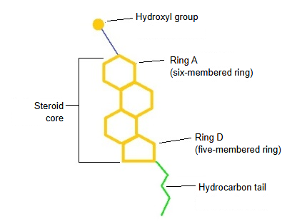
· Steroid core: a flat, rigid structure; it restricts the movement of the tails and regulates the stability and rigidity of the membrane.
· Hydroxyl group: hydrophilic; it interacts with the region near the phospholipid heads and acts as an anchor that fixes the position of the cholesterol.
· Hydrocarbon chain: hydrophobic; it intertwines with the tails and stabilises and fine-tunes the inside of the membrane.
⟨ Main functions ⟩
1. Regulating membrane fluidity: at high temperature it keeps the lipids from becoming too soft; at low temperature it keeps them from becoming too rigid.
2. Stabilising the membrane: it "links" the lipid bilayer and adjusts the thickness and rigidity of the membrane.
3. Supporting protein function: it sets up the local environment of transmembrane proteins and receptors and takes part in particular signalling (within lipid rafts).
4. Forming membrane microdomains (lipid rafts): together with sphingolipids it forms special domains of the membrane, which serve as platforms for signalling and membrane transport.
Fluidity
The cell membrane is fluid: its lipids and membrane proteins are moving.
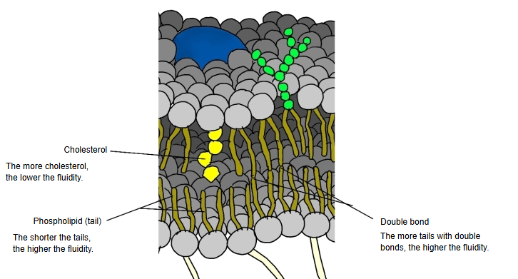
· The shorter the phospholipid tails (hydrocarbon chains), the higher the fluidity.
· At body temperature, the more cholesterol in the membrane, the lower the fluidity.
· The more tails with double bonds, the higher the fluidity. A double bond here is a carbon-carbon double bond (C=C) within one fatty-acid tail (an unsaturated fatty acid); it puts a kink in that tail, so the lipids cannot pack tightly.
Not all membrane proteins move freely.
Q: Are the phospholipids of the membrane constantly renewed?
Q: Are the phospholipids that form the membrane constantly being renewed?
A: Yes. The phospholipids of the membrane are constantly renewed (made, broken down and exchanged), but the membrane itself is not broken down and rebuilt.
1. What is exchanged
Exchanged: the phospholipid molecules themselves, the make-up of their fatty-acid chains and, indirectly, the kinds of head groups. Not exchanged: the basic structure of the lipid bilayer and the continuity of the membrane. The membrane is not a fixed object but a structure that keeps its shape while its molecules flow and are replaced.
2. How dynamic it is
Sideways movement (lateral diffusion): phospholipids move freely and very quickly within the plane of the membrane. Renewal at the molecular level: synthesis in the endoplasmic reticulum on its cytoplasmic side, breakdown and rebuilding by phospholipases and other enzymes; the molecules are renewed over minutes to days.
3. How they are exchanged
Synthesis: mainly in the endoplasmic reticulum (ER), from where the lipids reach each membrane by vesicular transport and by lipid-transfer proteins. Breakdown and reuse: they are broken down by phospholipases, and the fatty acids and head groups are often reused.
4. Why renewal is needed
Membrane lipids are damaged by oxidation; the fatty-acid make-up is changed to adjust the fluidity of the membrane; and lipids are used in signalling (for example PI to PIP2). The membrane must be dynamic in order to work.
5. How the asymmetry is kept
Phospholipids do not move between the inner and outer layers on their own; flippases, floppases and scramblases control this movement, so the different lipid make-up of the two layers is kept even though the lipids are renewed.
Reference websites
[Wikipedia]
· Cell membrane, Wikipedia.
· Lipid bilayer, Wikipedia.
· Membrane fluidity, Wikipedia.
[Websites with illustrations]
· Cell membrane, detailed diagram (Wikimedia Commons)

· Phospholipid bilayer (Microbe Notes)