Anterior triangle of neck (Latin: trigonum cervicale anterius)
Also called the anterior cervical triangle or the anterior triangle
Overview
The smaller triangles in the anterior triangle: submental triangle, submandibular triangle, carotid triangle and muscular triangle.
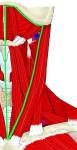 Anterior triangle
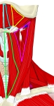 Smaller triangles
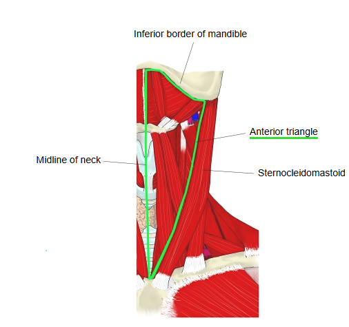
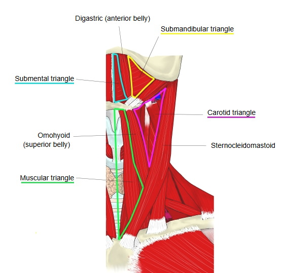
Below is a simple outline of the triangular depressions of the neck.
depressions
of the neck
- Anterior triangle : sternocleidomastoid (anterior border), midline of the neck, body of mandible (lower border)
- Submandibular triangle : digastric (posterior and anterior bellies), body of mandible
- Submental triangle : anterior bellies of the left and right digastric, hyoid bone
- Carotid triangle : sternocleidomastoid, digastric (posterior belly), omohyoid (superior belly)
- Muscular triangle : sternocleidomastoid (anterior border), omohyoid, midline of the neck = omotracheal triangle
- Posterior triangle : sternocleidomastoid (posterior border), trapezius (anterior border), clavicle (middle third) = lateral cervical region
- Occipital triangle : trapezius (anterior border), sternocleidomastoid (posterior border), omohyoid (inferior belly)
- Omoclavicular triangle : omohyoid (inferior belly), sternocleidomastoid (posterior border), clavicle (middle third)
The anterior triangle is bounded by the anterior border of sternocleidomastoid, the midline of the neck and the lower border of the body of the mandible. Its roof is the investing layer of the deep cervical fascia, which spans between the left and right sternocleidomastoid muscles; deep to it lie the pharynx, larynx and thyroid gland, and behind them the cervical vertebral column and the prevertebral muscles, covered by the prevertebral fascia. The digastric and the superior belly of omohyoid divide it into the submandibular, submental, carotid and muscular triangles.
Reference websites
[Wikipedia]
· Anterior triangle of the neck, Wikipedia.
[Websites with illustrations]
· Anterior triangle of the neck: boundaries, contents and subdivisions (TeachMeAnatomy)
· Muscles and labelled triangles of the neck (Wikimedia Commons)

Structure
The triangle is inverted: its base lies above, under the chin and jaw, and its apex points down to the sternum.
| Inferior boundary (apex) | The jugular notch of the manubrium of the sternum |
| Anterior boundary | The midline of the neck, from the chin to the jugular notch |
| Posterior boundary | The anterior border of sternocleidomastoid |
| Superior boundary (base) | The lower border of the body of the mandible and a line from the angle of the mandible to the mastoid process |
The investing layer of the deep cervical fascia forms the roof. In front of the deep floor (the vertebral column and prevertebral muscles, see above) lie the pharynx, larynx, trachea and thyroid gland in the visceral (pretracheal) layer of fascia; many texts describe these viscera as the floor of the triangle. The muscles in the triangle are:
· Suprahyoid muscles: digastric (anterior and posterior bellies), mylohyoid, geniohyoid and stylohyoid.
· Infrahyoid muscles: omohyoid, sternohyoid, sternothyroid and thyrohyoid.
Nerve supply of these muscles: the anterior belly of the digastric and mylohyoid by the mylohyoid nerve (from the mandibular nerve); the posterior belly of the digastric and stylohyoid by the facial nerve; geniohyoid and thyrohyoid by fibres of C1 that travel with the hypoglossal nerve; sternohyoid, sternothyroid and omohyoid by the ansa cervicalis. The anterior belly of the digastric develops from the first pharyngeal arch, the posterior belly from the second.
Related terms
- Submental triangle
- Carotid triangle
- Posterior triangle of neck
- Anterior cervical region
- Suprahyoid muscles
- Infrahyoid muscles
- Cervical fascia
References and sources
- Further reading: Anterior triangle of the neck, Wikipedia.
- Federative International Programme for Anatomical Terminology (FIPAT). Terminologia Anatomica, 2nd ed. (TA2), 2019.