Acinus (Latin: acinus glandulae)
Also called: glandular acinus (plural: acini)
- Overview
- Organs with acini
- Number of cells in an acinus
- Reference websites
- (Wikipedia, websites with illustrations)
- Related terms
Overview
- Exocrine cells : one kind of secretory cell; some form gland structures, most often an acinus, and some do not, such as the goblet cell.
- Lumen : the space inside the acinus, where the secretion is first released (acinar lumen).
- Duct : the route that carries the secretion away from the lumen; some duct cells also secrete or change the secretion.
cells
- Forming a gland : a group of exocrine cells + a duct.
- Acinar : end pieces shaped like a bunch of grapes; e.g. the pancreas, the parotid gland. The most common form.
- Tubular : long, narrow tubes; e.g. the intestinal glands of the large intestine, the gastric glands, the eccrine sweat glands. Fewer in number.
- Tubuloacinar : both tubes and acini; e.g. the submandibular gland, the sublingual gland.
- Alveolar (saccular) : rounder, sac-like end pieces, similar to acini; e.g. the mammary gland, the sebaceous glands.
- Not forming a gland : no duct: goblet cells, the Paneth cells of the intestine, the club (Clara) cells of the airway epithelium.
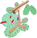 Acini and pancreatic islet
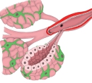 Mammary gland
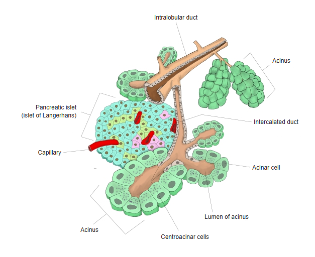
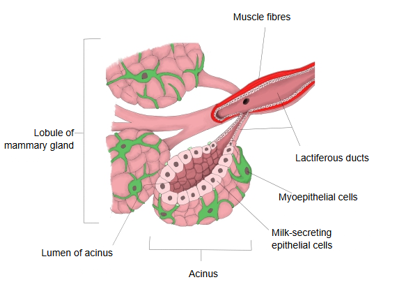
Organs with acini
| Organ | Type of acinus | Secretion | |
|---|---|---|---|
| 1 | Pancreas (exocrine part) | Serous acini | Digestive enzymes (amylase, lipase and others) |
| 2 | Parotid gland (salivary gland) | Serous acini | Serous saliva (contains digestive enzymes) |
| 3 | Submandibular gland (salivary gland) | Mixed acini (serous + mucous) | Mixed saliva |
| 4 | Sublingual gland (salivary gland) | Mainly mucous acini | Mucous saliva |
| 5 | Lacrimal gland | Serous acini | Tears (with lysozyme and others) |
| 6 | Mammary gland | Acinus-like alveoli (in the mammary lobules) | Milk (proteins, fats, lactose) |
| 7 | Sweat glands (eccrine glands) | Coiled tubular gland (no true acini) | Water, salts, urea and others |
| 8 | Sebaceous glands | Acinus-like (alveolar) end-pieces | Sebum (lipids) |
| 9 | Prostate | Tubuloalveolar (often called prostatic acini) | Prostatic fluid (part of semen) |
| 10 | Nasal glands and airway glands | Mixed (seromucous) acini, with many mucous cells | Mucus (mucins) and serous fluid |
Number of cells in an acinus
How many cells make up an acinus?
The number of exocrine cells in one acinus differs with the gland and the kind of acinus; the figures below are rough guides.
Typical number
Cells seen in one acinus in a section: about 5 to 20.
Examples
Acini of the pancreas (an exocrine gland)
· A section through one acinus shows roughly 8 to 12 acinar cells.
· The acinar cells are pyramid-shaped, with a broad base and a narrow apex, and release their secretion towards the centre of the acinus (the lumen).
Salivary glands such as the parotid gland and the submandibular gland
· A section through one acinus shows roughly 6 to 15 cells.
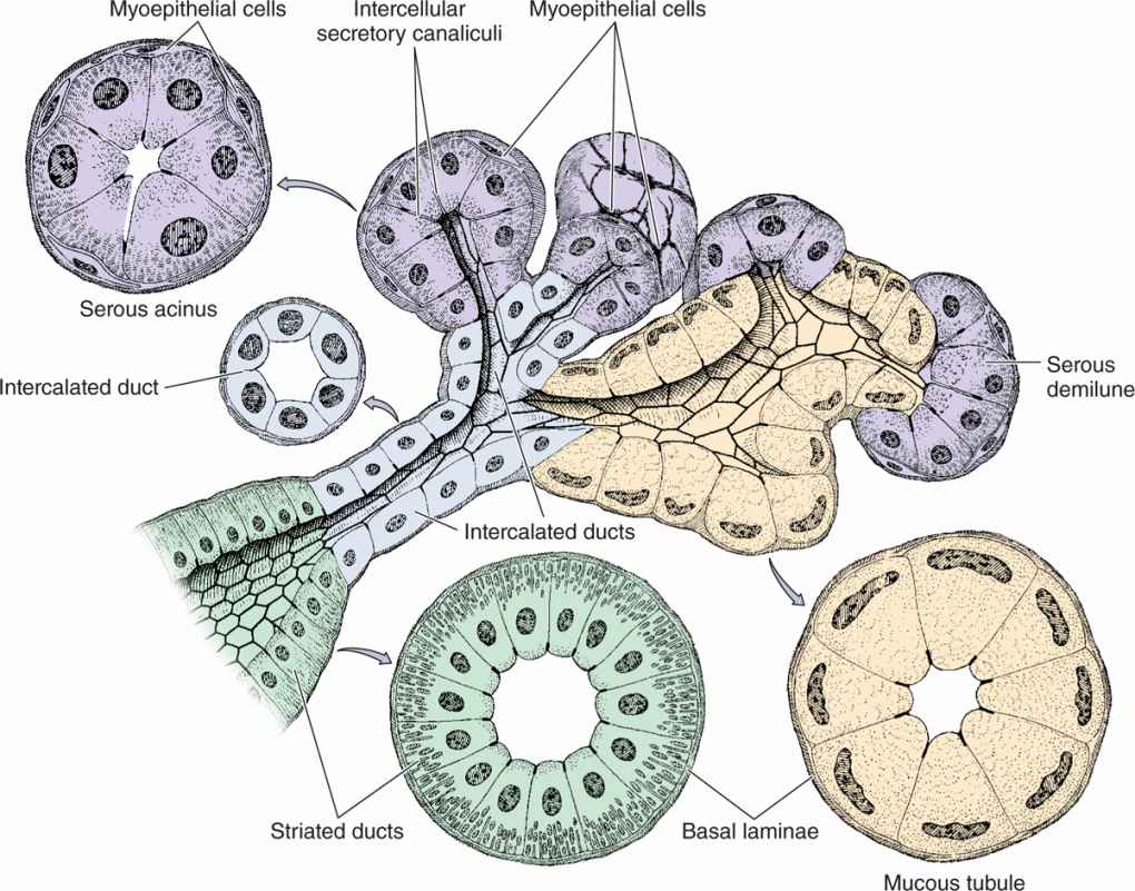
· Serous glands have serous acini and mucous glands have mucous acini; they differ somewhat in shape and cell number (mucous end-pieces are often larger and more tube-like).
Note
A section cuts through only part of an acinus, so the whole number of cells cannot be seen at once; histology texts therefore give a range of a few to a dozen or more cells per profile. The complete three-dimensional acinus contains several dozen cells (sources differ).
The cells are arranged around the acinar lumen; the secretion is released into it and then flows on into the ducts, starting with the intercalated duct.
Reference websites
[Wikipedia]
· Acinus, Wikipedia.
· Pancreatic acinar cell, Wikipedia.
[Websites with illustrations]
· Salivary gland acini and ducts (figure) (Pocket Dentistry)
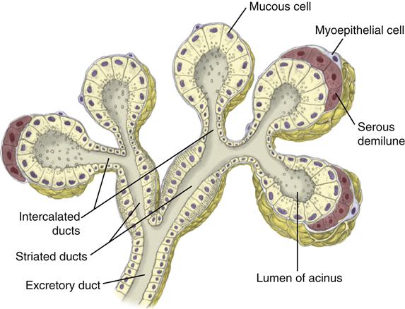
· Serous acinus, mucous tubule with serous demilune and the duct system (drawing) (Basicmedical Key)
Related terms
References and sources
- Further reading: Acinus, Wikipedia.
- Federative International Programme for Anatomical Terminology (FIPAT). Terminologia Anatomica, 2nd ed. (TA2), 2019.