Iliotibial tract
Also called the iliotibial band (IT band) or Maissiat's band.
Overview
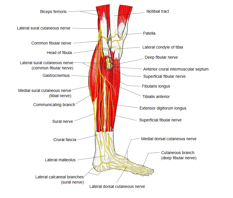
It is a band in which the fibres are packed very densely and strongly; its lower end is attached to the lateral condyle of the tibia.
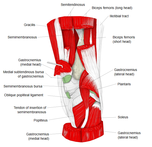
· Deep to it lies vastus lateralis. From its deep surface the lateral intermuscular septum passes to the linea aspera of the femur, separating vastus lateralis from the short head of biceps femoris.
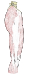 Fasciae of the lower limb
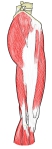 Muscles inserting into it
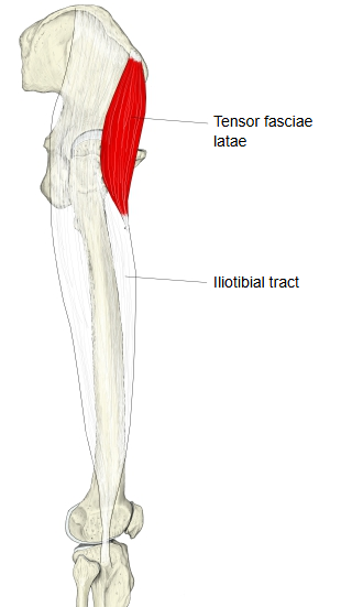
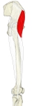 Tensor fasciae latae
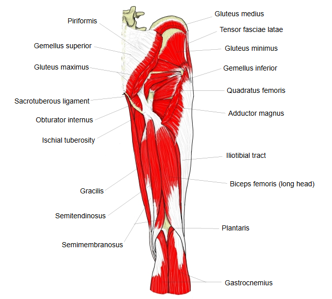
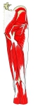 Hip and thigh (posterior view)
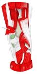 Back of the knee
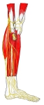 Leg (lateral view)
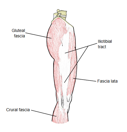
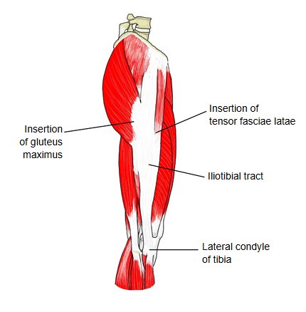
More detail
The iliotibial tract is a long, thick band of the fascia lata on the lateral side of the thigh. It begins at the iliac tubercle on the outer lip of the iliac crest and ends on the anterolateral surface of the lateral condyle of the tibia, at Gerdy's tubercle; some fibres also reach the patella. Tensor fasciae latae and much of the superficial part of gluteus maximus insert into it, so the tract acts as their common tendon. Above, it splits to enclose tensor fasciae latae, and its deep layer there blends with the lateral part of the capsule of the hip joint.
Pulled by these muscles, the tract helps to steady the hip and the knee joint on the lateral side, for example when the opposite foot is lifted off the ground. With the knee straight its lower part lies in front of the lateral epicondyle of the lateral condyle of the femur; with the knee bent beyond about 30 degrees it lies behind it (some authors think this shift only reflects changing tension in its front and back fibres). Repeated movement over the lateral femoral epicondyle, mainly in runners and cyclists, can cause pain on the outer side of the knee (iliotibial band syndrome).
Reference websites
[Websites with illustrations]
· Iliotibial band (Radiopaedia)
Related terms
- Fascia lata
- Deep fascia
- Lateral intermuscular septum
- Tensor fasciae latae
- Gluteus maximus
- Vastus lateralis
- Biceps femoris
- Iliac crest
- Lateral condyle of tibia
- Tibia
- Femur
- Hip joint
- Knee joint
References and sources
- Iliotibial tract, Wikipedia.
- FIPAT. Terminologia Anatomica, 2nd ed. (TA2). Federative International Programme for Anatomical Terminology, 2019: iliotibial tract (tractus iliotibialis), TA2 2690.