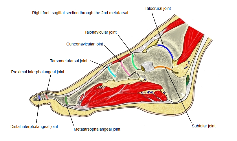
Talocrural joint (Latin: articulatio talocruralis)
Also called the ankle joint or the upper ankle joint
Overview
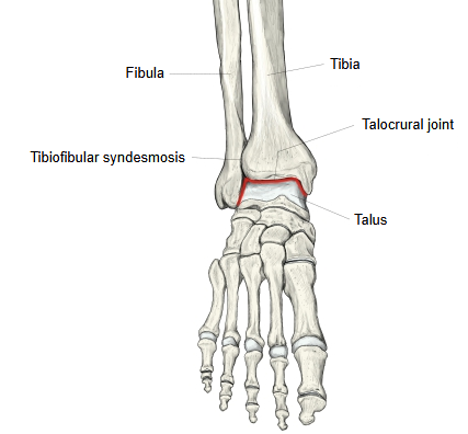
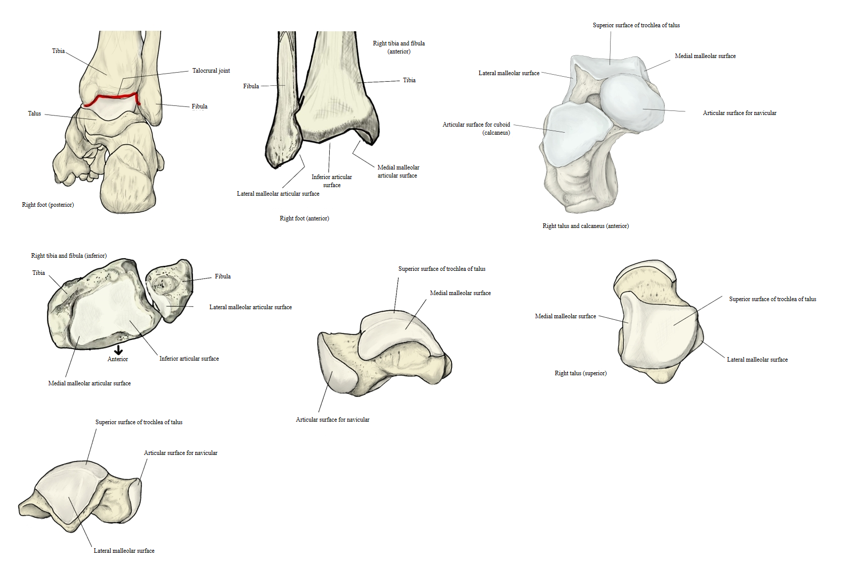
One of the joints of the foot: the joint of the ankle itself. The trochlea of the talus forms the articular head; the socket is formed by the inferior articular surface and the medial malleolar articular surface of the tibia, together with the lateral malleolar articular surface of the fibula. It is a spiral (cochlear) joint, a variety of hinge joint.
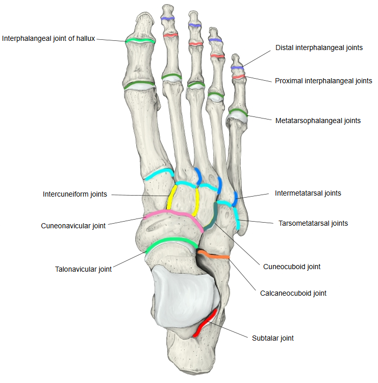
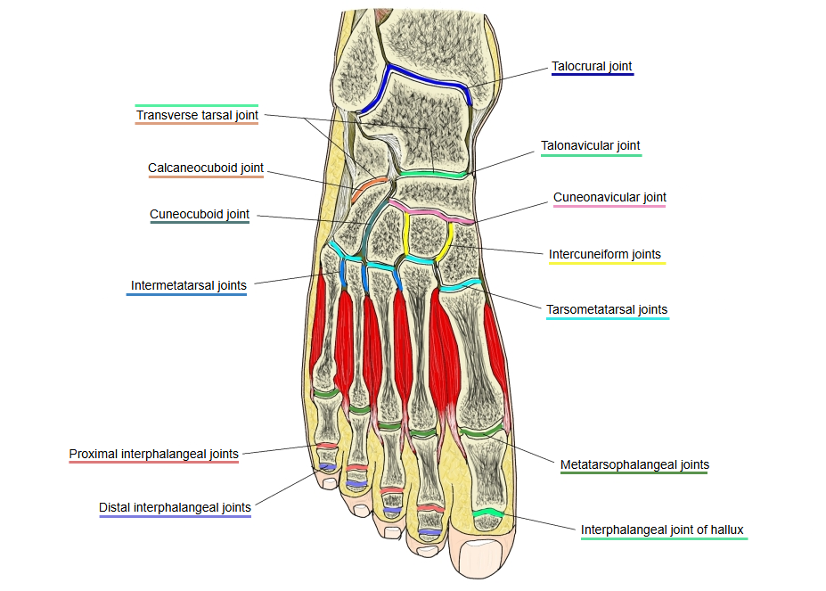
Joints of the foot
- Joints of the tarsus (2 kinds) : talocrural joint intertarsal joints (6 kinds)
- Joints of the metatarsus (2 kinds) : tarsometatarsal and intermetatarsal joints
- Joints of the toes (2 kinds) : metatarsophalangeal and interphalangeal joints
The talocrural, subtalar and talocalcaneonavicular joints work together when the foot pushes off, as in jumping, and are therefore also called the ankle joints (German Sprunggelenke):
| Joint | Also called |
|---|---|
| Talocrural joint | Upper ankle joint |
| Subtalar joint | Posterior lower ankle joint |
| Talocalcaneonavicular joint | Anterior lower ankle joint |
Bones
Talocrural joint: tibia (inferior articular surface, medial malleolar articular surface) and fibula (lateral malleolar articular surface) ⇔ talus (trochlea)
Type
Spiral (cochlear) joint — a hinge joint whose axis is slightly oblique
Joint capsule and movements
- The joint capsule arises from the margins of the articular cartilage and reaches only a little beyond the trochlea onto the neck of the talus. It is strong on the medial and lateral sides, where the collateral ligaments lie, and thin and loose in front and behind.
- The movements are mainly dorsiflexion and plantar flexion of the foot, as in a hinge joint; plantar flexion has the larger range.
- The trochlea of the talus, and the socket that holds it, are wider in front than behind. In dorsiflexion the wide front part is wedged into the socket and the joint is stable; in plantar flexion the narrow back part lies in the socket, and slight side-to-side and rotary movements of the foot become possible.
Shape of the articular surfaces
- The articular surface on the lower end of the tibia slopes slightly downward and medially and is concave from front to back. A low ridge running from front to back on this surface fits into a matching groove on the upper surface of the talus.
- The articular surface of the lateral malleolus (fibula) reaches further down than that of the medial malleolus (tibia).
- In sagittal section the trochlea of the talus is almost a circular arc, and its arc is larger than that of the tibial surface. Its medial edge runs straight forward, while its lateral edge runs forward and laterally; this is why the trochlea is wider in front. The lateral edge is also higher than the medial edge.
- The joint cavity is separate from those of the other joints of the foot; it extends a short way up into the narrow gap between the tibia and the fibula. A transverse synovial fold projects from the front and from the back wall of the capsule.
 Right foot (dorsal view)
 Right foot (section)
 Right foot (sagittal section)
Ligaments
| Ligament | |
|---|---|
| 1 | Medial (deltoid) ligament |
| 2 | Anterior talofibular ligament |
| 3 | Posterior talofibular ligament |
| 4 | Calcaneofibular ligament |