Latin: vertebrae thoracicae · T1–T12
Overview
- The twelve thoracic vertebrae lie below the cervical vertebrae; they are numbered from above, T1 to T12.
- Unlike the cervical vertebrae, their bodies carry costal facets for the heads of the ribs, and most of their transverse processes carry costal facets for the tubercles of the ribs.
- They have the typical form of a vertebra: a vertebral body, a vertebral arch and processes.
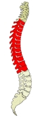
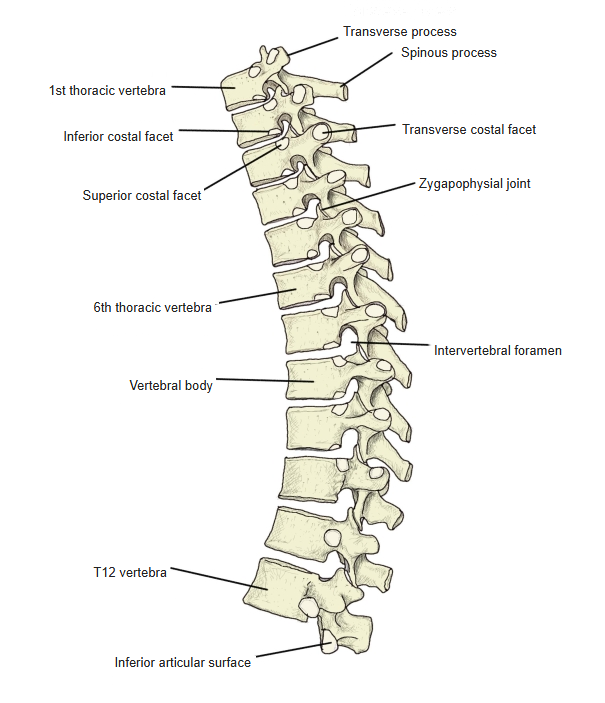
Thoracic vertebrae in the vertebral column (lateral) 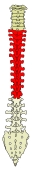
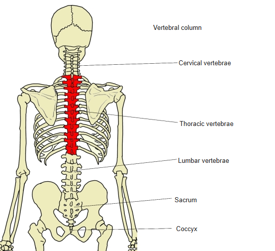
Thoracic vertebrae in the vertebral column (posterior) 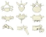
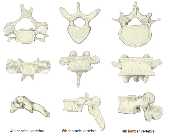
Cervical, thoracic and lumbar vertebrae compared
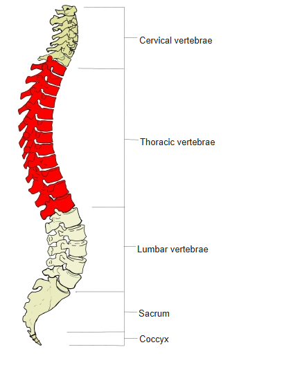
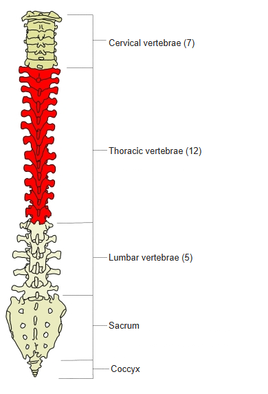
- Bones of the trunk
- Vertebral column
- Cervical vertebrae (7)
- Thoracic vertebrae (12)
- Lumbar vertebrae (5)
- Sacrum (1)
- Coccyx (1)
- Thoracic cage
- Sternum (1)
- Ribs (24)
- Vertebral column
Illustrations
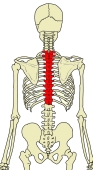
Position of the thoracic vertebrae 
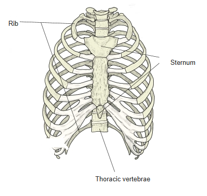
Thorax (anterior) 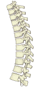
Thoracic vertebrae (left lateral) 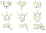
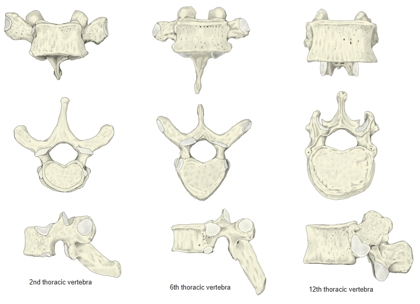
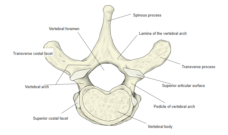
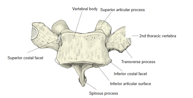
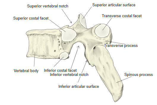
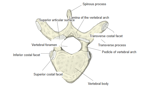
2nd, 6th and 12th thoracic vertebrae 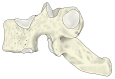
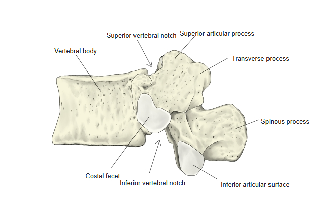
2nd thoracic vertebra (left lateral) 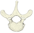
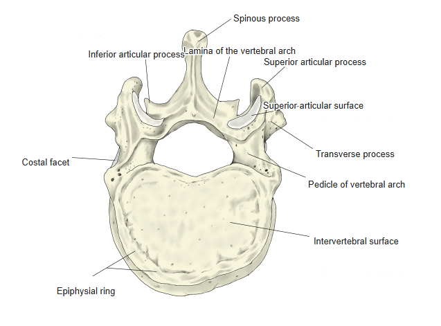
2nd thoracic vertebra (from above) 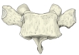
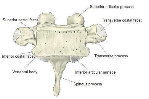
2nd thoracic vertebra (anterior) 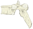
6th thoracic vertebra (left lateral) 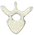
6th thoracic vertebra (from above) 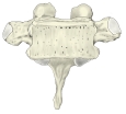
6th thoracic vertebra (anterior) 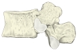
12th thoracic vertebra (left lateral) 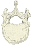
12th thoracic vertebra (from above) 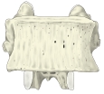
12th thoracic vertebra (anterior)
Articulations
| # | Bone | Joint |
|---|---|---|
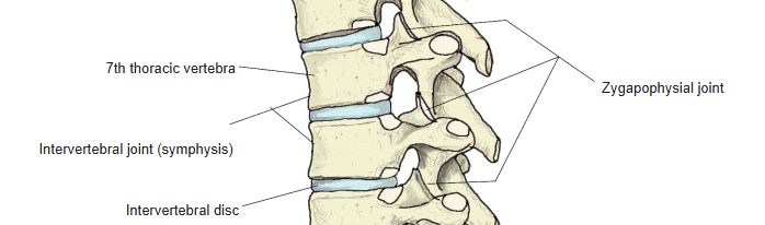
| 1 | Inferior articular surface ⇔ superior articular surface of the vertebra below | Zygapophysial (facet) joints |
| 2 | Vertebral bodies (through the intervertebral discs) | Intervertebral joints (symphyses) |
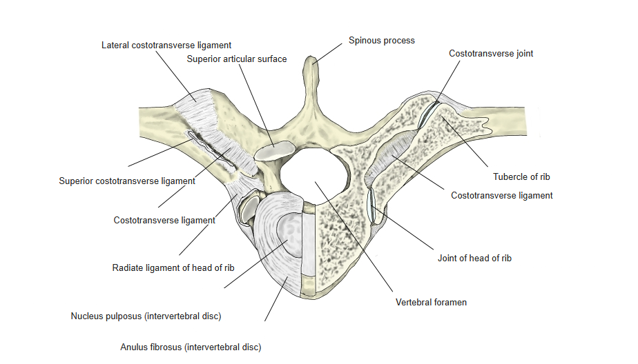
| 3 | Transverse process ⇔ tubercle of rib | Costotransverse joint |
| 4 | Costal facets ⇔ head of rib | Joint of head of rib |
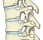
Joints between thoracic vertebrae 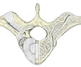
Joints with the ribs
Ligaments
The following ligaments attach to the thoracic vertebrae. The list is intended to be comprehensive but may omit minor ligaments.
| # | Ligament | Connects |
|---|---|---|
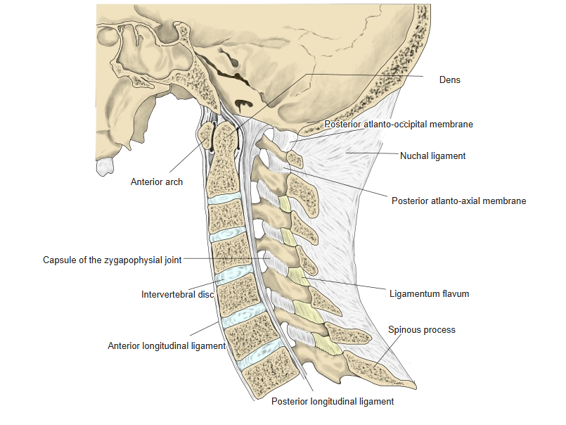
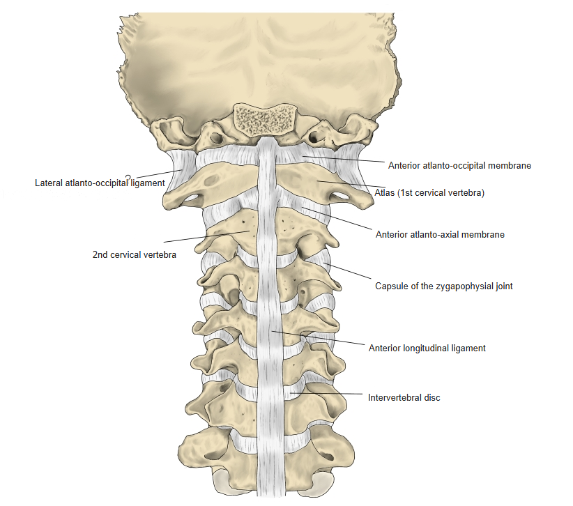
| 1 | Anterior longitudinal ligament | Occipital bone (pharyngeal tubercle) – front of the sacrum |
| 2 | Intertransverse ligaments | Between the transverse processes |
| 3 | Posterior longitudinal ligament | Occipital bone (clivus) – sacral canal |
| 4 | Ligamenta flava | Between the laminae |
| 5 | Interspinous ligaments | Between the spinous processes |
| 6 | Supraspinous ligament | Tips of the spinous processes |
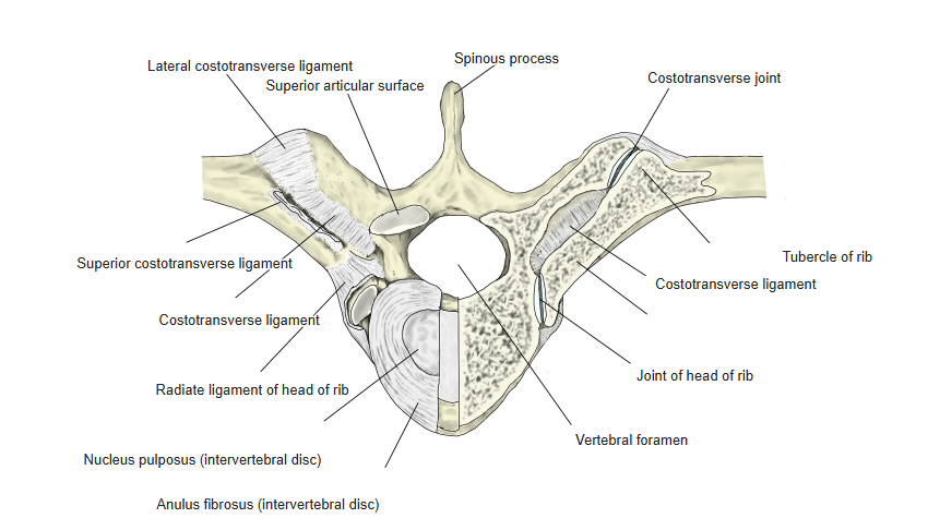
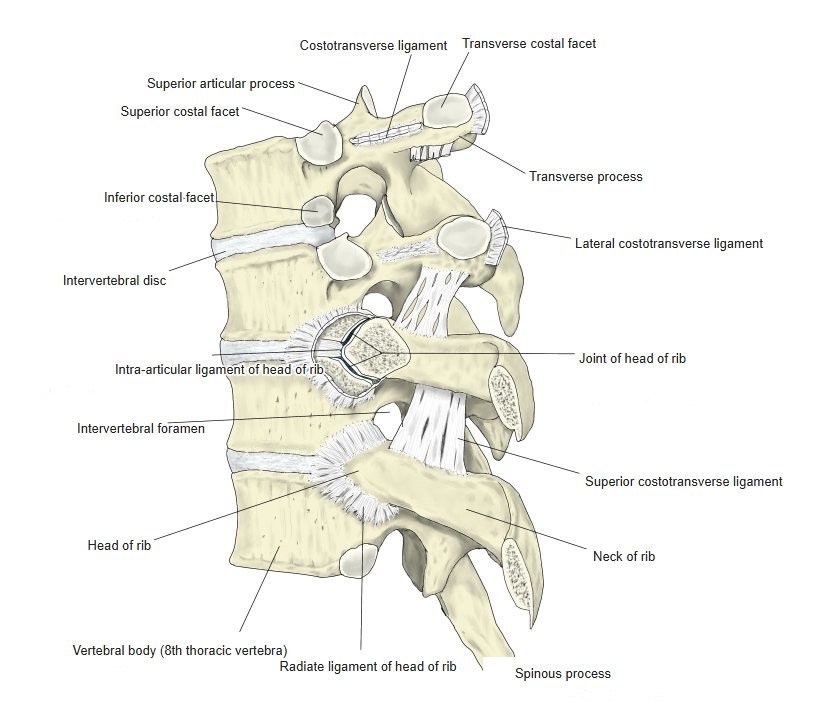
| 7 | Lateral costotransverse ligament | Transverse process – tubercle of rib |
| 8 | Superior costotransverse ligament | Transverse process – neck of the rib below |
| 9 | Costotransverse ligament | Transverse process – neck of rib |
| 10 | Radiate ligament of head of rib | Vertebral bodies – head of rib |
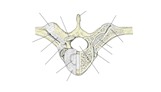
Costovertebral ligaments (from above) 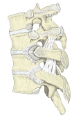
Costovertebral ligaments (lateral) 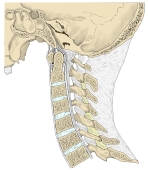
Ligaments of the cervical spine (median section) 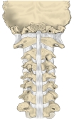
Ligaments of the cervical spine (anterior)
Muscle attachments
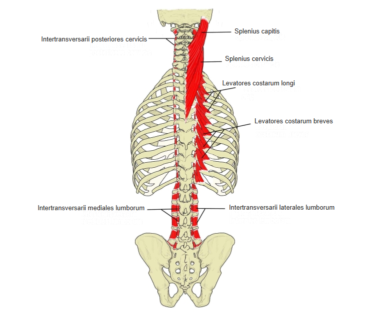
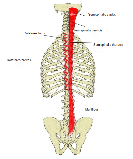
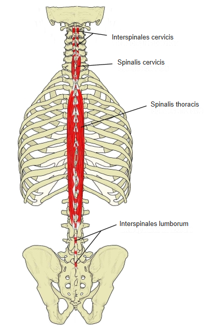
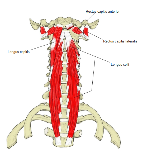
- Origin of: Trapezius, Latissimus dorsi, Rhomboid major, Serratus posterior superior, Splenius capitis, Splenius cervicis, Spinalis cervicis, Interspinales thoracis, Longissimus, Semispinalis, Rotatores, Longus colli, Intertransversarii, Rhomboid minor, Serratus posterior inferior, Spinalis thoracis, Multifidus
- Insertion of: none (all muscles that insert here also arise here and are listed under origin)
Muscles that both arise from and insert on thoracic vertebrae are listed under origin.
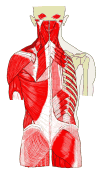
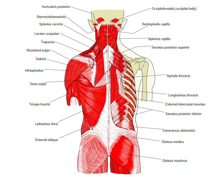
Muscles of the back (superficial and deep) 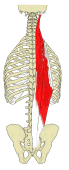
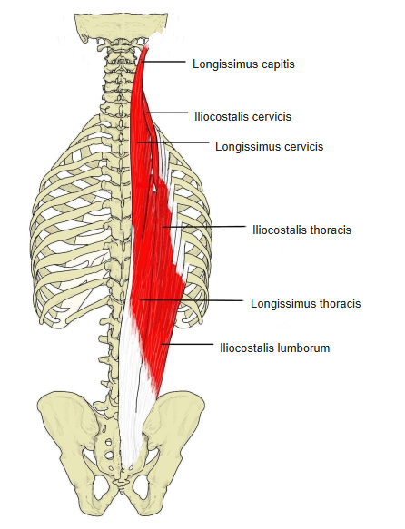
Erector spinae 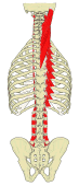
Splenius and intertransverse muscles 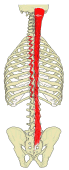
Transversospinales 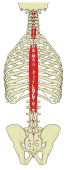
Spinalis and interspinales 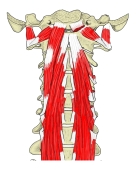
Prevertebral muscles (anterior)
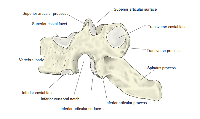
Parts of a thoracic vertebra
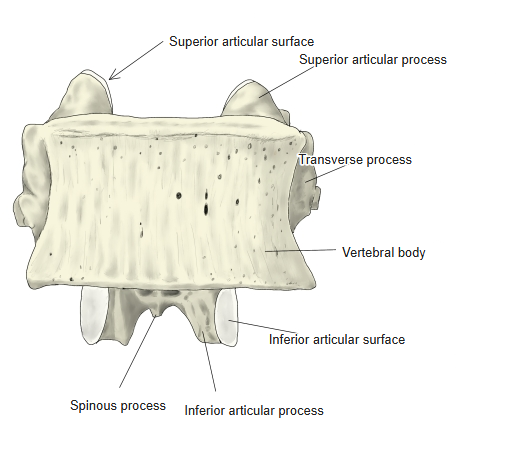
- Vertebral body – larger than in the cervical vertebrae but smaller than in the lumbar vertebrae; the bodies increase in size from T1 to T12. Its sides carry the superior and inferior costal facets (demifacets) for the heads of the ribs.
- Transverse process – best developed at T7 or T8. Its tip carries the transverse costal facet for the tubercle of a rib (not on T11 and T12). Unlike the cervical vertebrae, it has no anterior or posterior tubercle and no groove for the spinal nerve.
- Spinous process – long and pointing steeply downwards, overlapping the vertebra below in the middle of the thoracic region.
- Vertebral foramen – almost round, and much smaller than in the cervical vertebrae.
References and sources
- Further reading: Thoracic vertebrae, Wikipedia.