Latin: os temporale
Overview
- A paired bone forming part of the side and base of the skull; it houses the middle and inner ear.
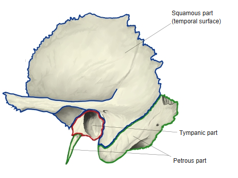
- It has three main parts: the squamous part, the tympanic part and the petrous part (including the mastoid region); some texts also count the styloid region separately. The parts fuse into one bone about a year after birth.
- It meets four bones by sutures: the parietal, occipital, sphenoid and zygomatic bones. It also articulates with the mandible at the temporomandibular joint (a synovial joint, not a suture).
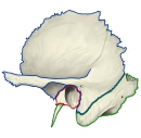
Parts of the temporal bone
- Bones of the skull
- Neurocranium (cranial bones)
- Frontal bone
- Parietal bone (2)
- Occipital bone
- Temporal bone (2)
- Sphenoid bone
- Ethmoid bone
- Viscerocranium (facial bones)
- Nasal bone (2)
- Vomer
- Lacrimal bone (2)
- Inferior nasal concha (2)
- Maxilla (2)
- Zygomatic bone (2)
- Palatine bone (2)
- Mandible
- Hyoid bone
- Neurocranium (cranial bones)
Illustrations
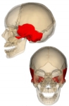
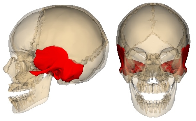
Position of the temporal bone 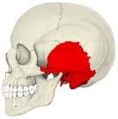
Skull (left lateral) 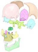
Bones of the skull (separated) 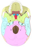
External cranial base 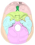
Internal cranial base
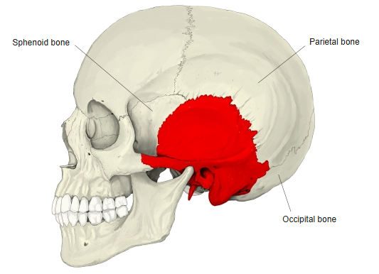
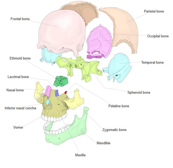
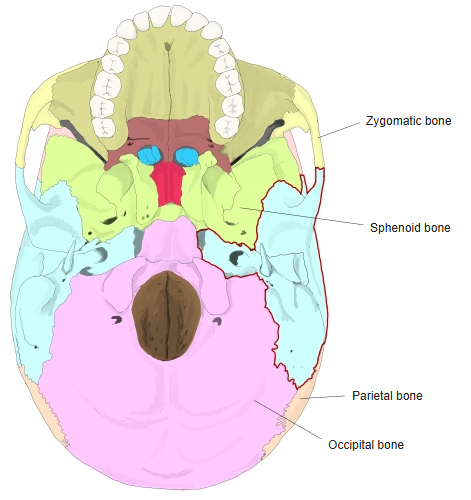
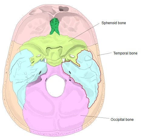
Articulations
| # | Bone | Joint |
|---|---|---|
| 1 | Parietal bone | Squamous suture, parietomastoid suture |
| 2 | Occipital bone | Occipitomastoid suture, petro-occipital fissure |
| 3 | Sphenoid (greater wing) | Sphenosquamous suture, sphenopetrosal fissure |
| 4 | Zygomatic bone | Temporozygomatic suture |
| 5 | Mandible (head) | Temporomandibular joint |
Ligaments
The following ligaments attach to the temporal bone. The list is intended to be comprehensive but may omit minor ligaments.
| # | Ligament | Connects |
|---|---|---|
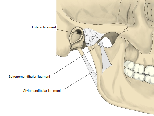
| 1 | Lateral ligament (temporomandibular joint) | Zygomatic arch and articular tubercle – neck of mandible |
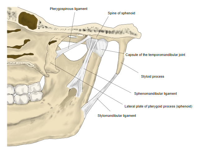
| 2 | Stylomandibular ligament | Styloid process – angle of mandible |
| 3 | Sphenomandibular ligament | Spine of sphenoid – lingula of mandible (passes close to the temporal bone) |
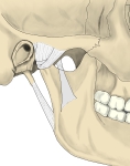
Ligaments of the temporomandibular joint (lateral) 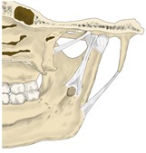
Ligaments of the temporomandibular joint (medial)
Muscle attachments
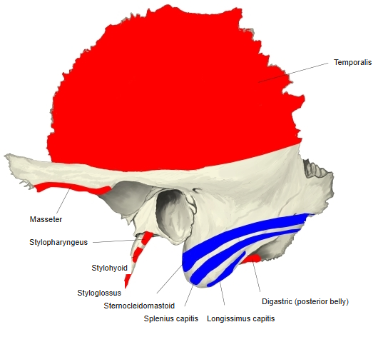
- Origin of: Temporalis, Masseter, Stylohyoid, Styloglossus, Stylopharyngeus, Levator veli palatini, Digastric (posterior belly)
- Insertion of: Sternocleidomastoid, Splenius capitis, Longissimus capitis
Masseter (deep part) arises from the zygomatic process; the three styloid muscles from the styloid process; digastric from the mastoid notch; sternocleidomastoid, splenius capitis and longissimus capitis insert on the mastoid process.
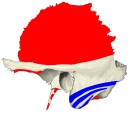
Muscle attachments (lateral) 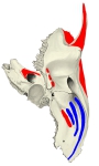
Muscle attachments (from below)
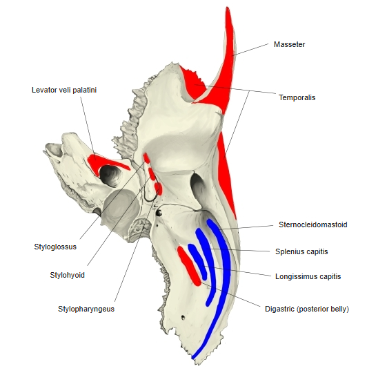
Borders
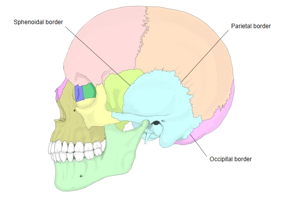
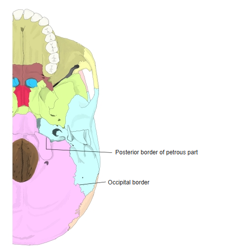
- Occipital border – meets the occipital bone behind.
- Posterior border of the petrous part – its front-medial half meets the basilar part of the occipital bone to form the petro-occipital fissure.
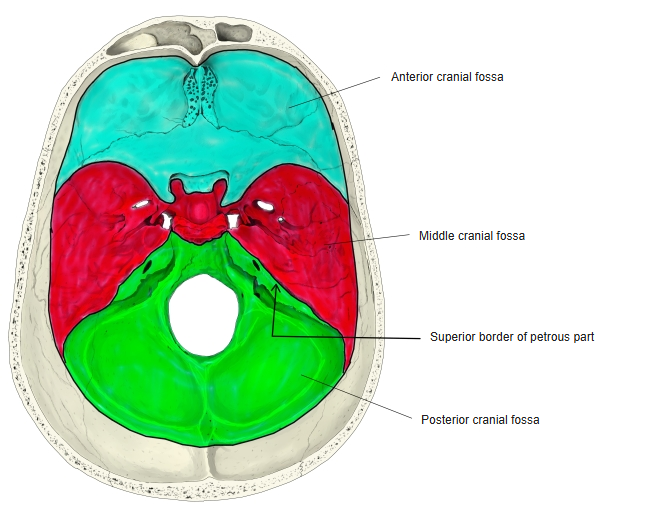
- Superior border of the petrous part – a sharp ridge separating the middle and posterior cranial fossae; it carries the groove for the superior petrosal sinus.
- Sphenoidal border – a serrated edge meeting the greater wing of the sphenoid in front.
- Parietal border – behind the sphenoidal border; meets the parietal bone.
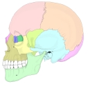
Borders (lateral view) 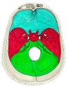
Cranial fossae 
Borders (external cranial base)
Surfaces
- The squamous part has an outer (temporal) and an inner (cerebral) surface.
- The petrous part is usually described with four surfaces (anterior, posterior and inferior surfaces, and the base).
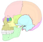
Skull (left lateral, named) 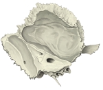
Inner surfaces 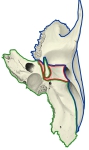
Inferior surface
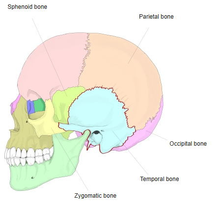
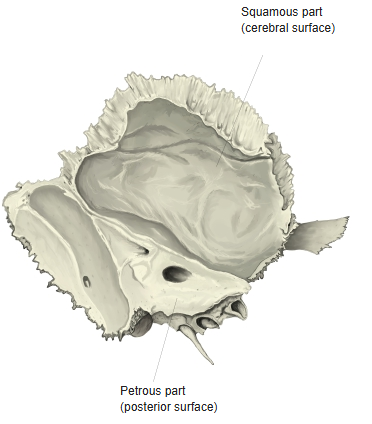
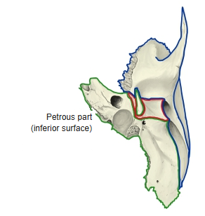
Foramina and openings
- External acoustic opening – the entrance of the external acoustic meatus.
- Internal acoustic opening – facial, intermediate and vestibulocochlear nerves; labyrinthine artery and veins.
- Stylomastoid foramen – facial nerve; stylomastoid artery.
- Jugular foramen (with the occipital bone) – glossopharyngeal, vagus and accessory nerves; internal jugular vein, inferior petrosal sinus; posterior meningeal artery.
- External and internal openings of the carotid canal – the entrance and exit of the carotid canal (internal carotid artery).
- Mastoid foramen – mastoid emissary vein.
- External opening of the cochlear canaliculus, openings of the tympanic canaliculus, external opening of the vestibular aqueduct.
- Tympanomastoid fissure – the tympanic canaliculus opens here; hiatus for the lesser petrosal nerve – at the back end of its groove.
References and sources
- Further reading: Temporal bone, Wikipedia.