Tibial tuberosity (Latin: tuberositas tibiae)
Also called the tibial tubercle
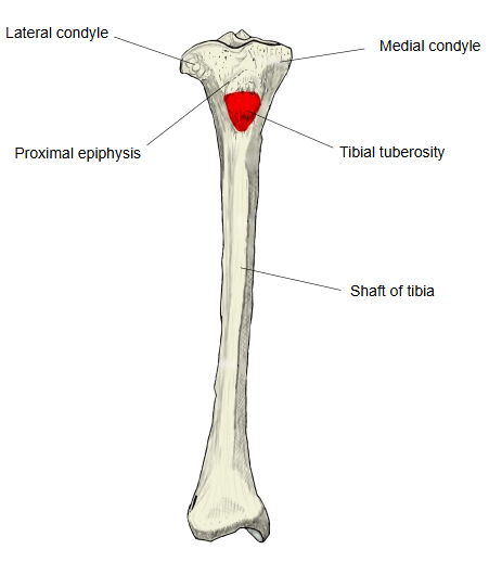
A large, rough elevation on the front of the upper end of the tibia, just below the condyles; the patellar ligament is attached to it.
It lies at the upper end of the anterior border of the shaft, below the front of the lateral and medial condyles. Its smoother upper part receives the patellar ligament; its lower, rougher part lies just under the skin. Through the patellar ligament it is the final attachment of quadriceps femoris (rectus femoris and the three vasti), so it takes the pull that extends the knee. It is easily felt below the kneecap and takes much of the weight in kneeling. In active adolescents, repeated pull of the patellar ligament can irritate the growing tuberosity (Osgood–Schlatter disease).
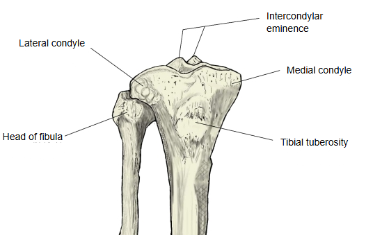
Attached structures
| Muscles | 1 | Vastus lateralis (insertion, via the patellar ligament) | musculus vastus lateralis | Right tibia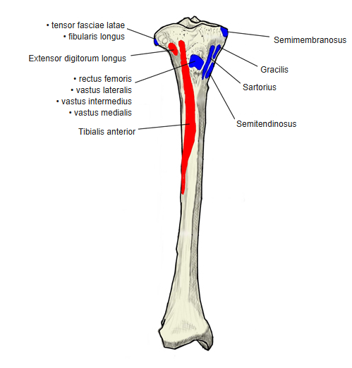 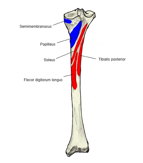 |
| 2 | Rectus femoris (insertion, via the patellar ligament) | musculus rectus femoris | ||
| 3 | Vastus intermedius (insertion, via the patellar ligament) | musculus vastus intermedius | ||
| 4 | Vastus medialis (insertion, via the patellar ligament) | musculus vastus medialis | ||
| 5 | Semitendinosus (insertion, medial to the tuberosity) | musculus semitendinosus | ||
| 6 | Sartorius (insertion, medial to the tuberosity) | musculus sartorius | ||
| 7 | Gracilis (insertion, medial to the tuberosity) | musculus gracilis | ||
| Ligament | 1 | Patellar ligament | ligamentum patellae |
The tendons of sartorius, gracilis and semitendinosus insert close by, on the medial surface just medial to the tuberosity (pes anserinus), not on the tuberosity itself.
Reference websites
[Wikipedia]
· Tuberosity of the tibia, Wikipedia.
· Osgood–Schlatter disease, Wikipedia.
[Websites with illustrations]
· Bones of the right leg, anterior surface (Gray's Anatomy, fig. 258) (Wikimedia Commons)

· The tibia (TeachMeAnatomy)
· Tibia (Kenhub)
Where it is found
Related terms
- Anterior border of tibia
- Lateral condyle of tibia
- Medial condyle of tibia
- Proximal epiphysis of tibia
- Shaft of tibia
- Knee joint
References and sources
- Further reading: Tuberosity of the tibia, Wikipedia.
- Further reading: Osgood–Schlatter disease, Wikipedia.
- Federative International Programme for Anatomical Terminology (FIPAT). Terminologia Anatomica, 2nd ed. (TA2), 2019.