Antitragicus (Latin: musculus antitragicus)
- Overview
- Illustrations
- Origin / insertion
- Action
- Nerve / blood supply
- Reference websites
- (Wikipedia, websites with illustrations)
- Related terms
Overview
- Facial muscles (24)
- Muscles of mastication (4)
- Extra-ocular muscles (7)
- Muscles of the auditory ossicles (2)
- Muscles of the tongue (7)
- Muscles of the soft palate and fauces (5)
· A small band running from the antitragus upward and backward to the tail of the helix.
· Like the other intrinsic auricular muscles it is small and weak; in most people it changes the shape of the auricle very little. These muscles develop from the second pharyngeal arch, which is why the facial nerve supplies them.
Illustrations
More figures: Multi-muscle illustrations: Head · Muscles of the head (list) · Auricle
 Left auricular cartilage (lateral surface)
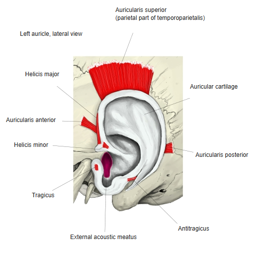
Origin / insertion
[Origin] : the outer (lateral) surface of the antitragus.
[Insertion] : the tail of the helix (cauda helicis) and the antihelix.
Action
Draws the antitragus and the tail of the helix toward each other, slightly changing the shape of the auricle; in some people this may widen the opening of the external acoustic meatus.
Nerve / blood supply
· Nerve : facial nerve (cranial nerve VII), temporal branches. Some texts add the posterior auricular nerve.
· Arteries : small branches of the posterior auricular artery and the anterior auricular branches of the superficial temporal artery.
· Veins : the posterior auricular vein and the anterior auricular veins.
Reference websites
[Wikipedia]
· Antitragicus, Wikipedia.
[Websites with illustrations]
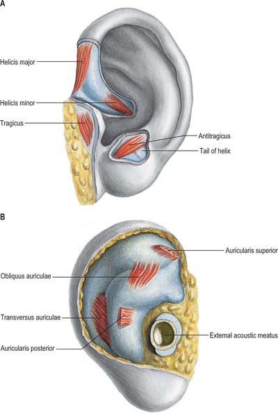
· Intrinsic muscles of the auricle (image) (Clinical Gate)
Related terms
- Auricular muscles
- Intrinsic auricular muscles
- Auricular cartilage
- Antitragus
- Cauda helicis
- Antihelix
- Tragicus
- Auricle
References and sources
- FIPAT. Terminologia Anatomica, 2nd ed. (TA2), 2019: musculus antitragicus.
- Antitragicus, Wikipedia.