Auricular muscles (Latin: musculi auriculares)
Also called the muscles of auricle
| Auricular muscles | Extrinsic | 1 | Auricularis anterior | Origin: epicranial aponeurosis. Insertion: front of the helix (auricular cartilage). | |
| 2 | Auricularis posterior | Origin: outer surface of the mastoid part of the temporal bone. Insertion: cranial (medial) surface of the auricular cartilage. | |||
| 3 | Auricularis superior | Origin: epicranial aponeurosis. Insertion: upper part of the cranial surface of the auricle. | |||
| Intrinsic | Lateral surface | 1 | Helicis major | Origin: spine of helix; runs up along the front border of the helix. | |
| 2 | Helicis minor | A small oblique band lying on the crus of helix. | |||
| 3 | Tragicus | Runs vertically on the outer surface of the tragus. | |||
| 4 | Antitragicus | Arises from the outer surface of the antitragus and is attached to the cauda helicis and the antihelix. | |||
| Cranial surface | 5 | Transverse muscle of auricle | Stretches between the eminence of concha and the eminence of scapha on the cranial (medial) surface of the auricle. | ||
| 6 | Oblique muscle of auricle | On the cranial surface, from the eminence of concha (the bulge behind the concha) to the eminence of triangular fossa above it. | |||
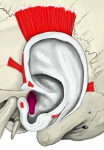 Left auricle (lateral surface)
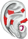 Left auricle (cranial surface)
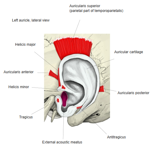
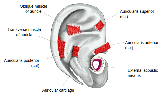
The auricular muscles belong to the muscles of facial expression and are supplied by branches of the facial nerve. In humans they are thin and mostly weak: auricularis anterior draws the ear forwards and upwards, auricularis superior raises it slightly and auricularis posterior draws it backwards, but few people can move their ears on purpose, and the intrinsic muscles barely change the shape of the auricle. The extrinsic muscles lie in the auricular region; for the cartilage they act on, see auricular cartilage.
Reference websites
[Wikipedia]
· Auricular muscles, Wikipedia.
[Websites with illustrations]
· Muscles of the auricle (figure I) (Springer Nature)

· Muscles of the auricle (figure II) (Springer Nature)

· Muscles of the auricle (atlas figure) (doctorlib.info)

Related terms
- Auricular tubercle
- Auricular cartilage
- Auricular region
- Concha of auricle
- Extrinsic auricular muscles
- Auricularis anterior
- Auricularis superior
- Auricularis posterior
- Auricle
- Epicranial aponeurosis
- Temporoparietalis
References and sources
- Further reading: Auricular muscles, Wikipedia.
- Federative International Programme for Anatomical Terminology (FIPAT). Terminologia Anatomica, 2nd ed. (TA2), 2019.