Enterocyte (Latin: enterocytus)
Also called Absorptive cell of the intestine or the intestinal absorptive cell
- Overview
- Functions
- Questions and answers
- Reference websites
- (Wikipedia, websites with illustrations)
- Where it is found
- Related terms
Overview
· Microvilli: length about 1 µm, diameter about 0.1 µm; about 1,000–2,000 or more per cell (estimates range from about one to several thousand). Together they form the brush border.
Neighbouring cells are sealed near the surface by tight junctions and held together by adherens junctions and desmosomes; their side walls interlock. The nucleus lies in the lower part of the cell, which rests on a basement membrane over the lamina propria, with its capillaries and central lacteal. Enterocytes arise from stem cells in the crypts, move up the villus and are shed at its tip after a few days, so the lining is renewed constantly.
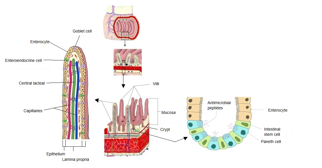
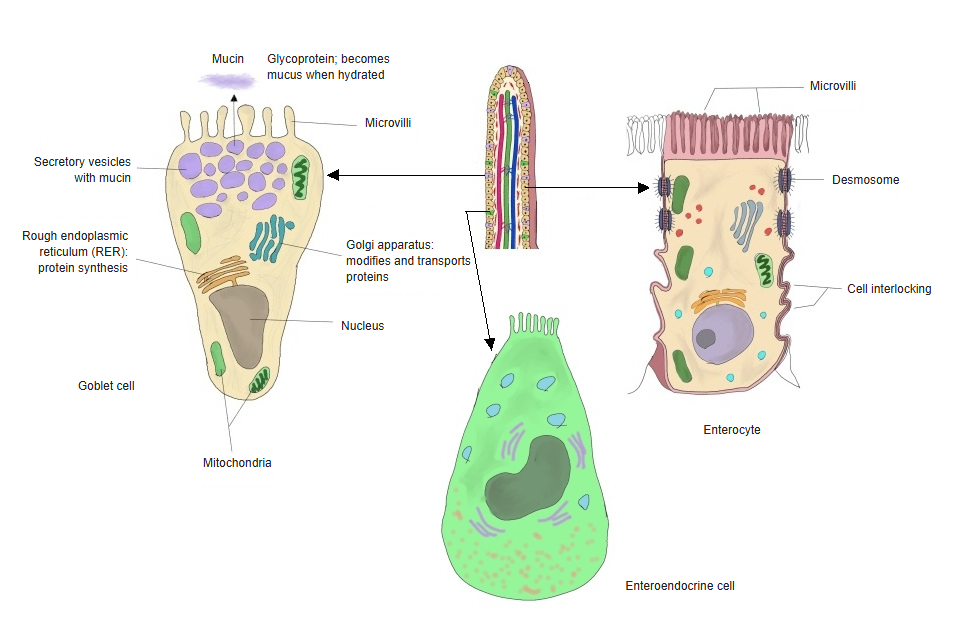
The table below is a short list of the epithelial cells of the mucosa of the small intestine.
| Cell type | Main function | Location | Share (approx.) | |
|---|---|---|---|---|
| 1 | Enterocytes | Absorption of nutrients; brush-border digestive enzymes | Villi and crypts | about 80–90% |
| 2 | Goblet cells | Secrete mucus | Villi and crypts | about 10–15% (varies by region; more towards the ileum) |
| 3 | Enteroendocrine cells | Secrete hormones | Mainly the crypts | about 1–2% |
| 4 | Paneth cells | Secrete antimicrobial peptides | Base of the crypts | about 1–3% (mainly in the jejunum and ileum) |
| 5 | Stem cells | Renew the epithelium | Base of the crypts | very few (<1%) |
Functions
1. 1. Absorption of nutrients
Sugars (such as glucose), amino acids and small peptides, fats (fatty acids and monoglycerides, packed into chylomicrons), vitamins, minerals and water. Sugars and amino acids pass into the capillaries and on to the portal vein; fats pass into the central lacteal.
2. 2. Digestive enzymes
The brush border membrane (the microvilli) carries built-in digestive enzymes, such as disaccharidases (maltase, sucrase, lactase) and peptidases, which complete digestion at the cell surface.
3. 3. Barrier function
The sealed sheet of cells keeps gut bacteria and harmful substances out of the tissues.
Questions and answers
Q: Do the enterocytes of the jejunum and the ileum differ?
A: Yes, in several points of structure and function. They are basically the same kind of cell, but their features are adapted to the nutrients absorbed and to immune functions.
1. 1. Development of the microvilli (brush border)
| Feature | Jejunum | Ileum |
|---|---|---|
| Height and density of the microvilli | Tall and dense | Lower and sparser |
| Membrane enzymes | Rich in digestive enzymes (maltase, sucrase and others) | Fewer digestive enzymes |
Meaning: the jejunum is specialized for absorbing carbohydrates and proteins, so its microvilli are long and dense to make absorption efficient.
The ileum mainly absorbs water, vitamin B12 and bile acids, so its microvilli are somewhat less developed.
2. 2. What is absorbed
Jejunum: active absorption of carbohydrates, amino acids and fats; rich in nutrient-splitting enzymes.
Ileum: adapted to absorb the vitamin B12–intrinsic factor complex and bile acids; fat absorption is less active than in the jejunum.
3. 3. Links with immune function
The ileum has many Peyer's patches (aggregated lymphoid nodules), and its enterocytes also express immune-related molecules (such as MHC class II and Toll-like receptors), taking part in catching antigens and in gut immunity.
In the jejunum, solitary lymphoid nodules predominate, and the immune function is weaker than in the ileum.
Q: Why are the borders between neighbouring cells jagged?
A: The contact surfaces of neighbouring enterocytes interlock in a jagged line, which strengthens their attachment. This is important for keeping up the barrier function of the intestinal tract.
1. 1. Structure of the contact surface
Neighbouring enterocytes do not meet along a simple flat plane: their surfaces form a zigzag of interlocking ridges and grooves.
Along these folds the cell membranes lie close together, and adhesion molecules can be placed effectively.
2. 2. Cell junctions
a. Tight junction (zonula occludens)
At the very top of the intestinal epithelium; seals the space between the cells.
Prevents leakage of water and solutes and secures the barrier function.
b. Desmosome
Found on the sides of the cells.
Binds the cells firmly together and withstands mechanical stress.
c. Adherens junction
Lies just below the tight junction.
Stabilizes the junction together with the cytoskeleton (actin).
3. 3. Advantages of the interlocking (zigzag) borders
Larger contact area → stronger attachment between the cells
Better barrier function → keeps harmful substances and pathogens in the gut from entering
Greater mechanical strength → withstands the friction and pressure of peristalsis
Q: What is the basement membrane under the enterocytes like?
A: The basement membrane under the enterocytes plays a very important part in supporting the cells, in their differentiation and in the barrier function. In outline:
1. 1. Position and structure
Position: between the base of the enterocytes and the connective tissue of the intestinal mucosa (the lamina propria).
Structure: a thin, fibrous sheet, about 50–100 nm thick under the electron microscope.
Main components:
Laminin: important for cell adhesion and for the signals of differentiation
Type IV collagen: forms the framework of the basement membrane
Heparan sulfate proteoglycans: hold extracellular signalling molecules
Nidogen: links laminin and type IV collagen
2. 2. Main functions
a. Support
Anchors the enterocytes firmly, so that they withstand the mechanical stress of the intestinal tract (such as peristalsis).
b. Cell adhesion and polarity
Adhesion molecules (such as integrins) that bind the basement membrane keep the polarity of the enterocytes (the difference between the apical and basal sides).
c. Control of differentiation and proliferation
Provides signals needed for the differentiation of stem cells into mature enterocytes.
d. Regulation of exchange
Regulates the passage of nutrients and other substances between the enterocytes and the blood and lymph vessels.
It also acts as a barrier against the passage of too much material.
3. 3. Related structures
· Contact with the cell base
The base of the cell rests closely on the basement membrane and is anchored to it, which supports attachment and exchange.
· Blood and lymph vessels
Beneath the basement membrane lies the connective tissue of the lamina propria with its capillaries and central lacteal; absorbed substances cross the basement membrane to reach them.
Reference websites
[Wikipedia]
· Enterocyte, Wikipedia.
[Websites with illustrations]
· Enterocyte (diagram) (Wikimedia Commons)

· Domains of the enterocyte (Atlas of Plant and Animal Histology, University of Vigo)

· Structure of the intestinal epithelium and enterocytes (Nutrixeal Info)

Where it is found
Related terms
References and sources
- Further reading: Enterocyte, Wikipedia.
- Federative International Programme for Anatomical Terminology (FIPAT). Terminologia Anatomica, 2nd ed. (TA2), 2019.