Inferior orbital fissure (Latin: fissura orbitalis inferior)
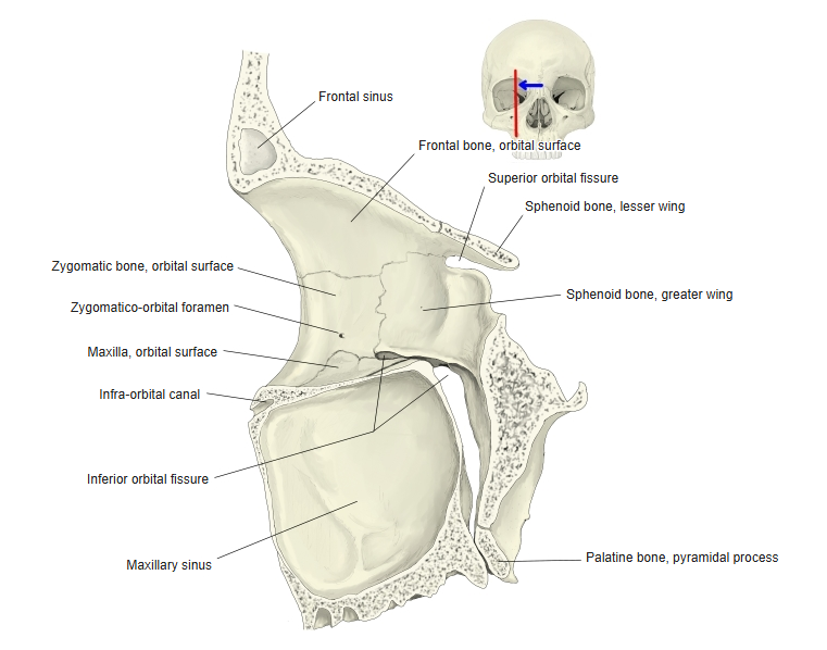
A slit that runs crosswise at the back of the lower part of the orbit, between its floor and its lateral wall. Nerves and vessels pass through it, and it connects the orbit with the infratemporal fossa and the pterygopalatine fossa.
Orbit ⇔ Inferior orbital fissure ⇔ Infratemporal fossa and pterygopalatine fossa
More details
[Bones forming it]
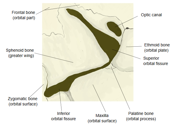
It runs forwards and laterally from the apex of the orbit, below and diverging from the superior orbital fissure. Behind, it opens into the pterygopalatine fossa; further forwards and laterally, into the infratemporal fossa. It is formed mainly by the following bones.
Below: the orbital surface of the maxilla.
Lateral end: the orbital surface of the zygomatic bone, only a small part, which often closes the end of the fissure.
The orbital process of the palatine bone may also be counted as a boundary at the back; its share is small and varies between skulls, so sources differ.
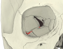 Right orbit (anterior view)
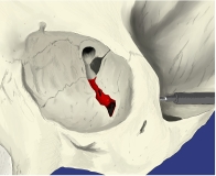 Left orbit (from above and in front)
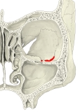 Orbits and nasal cavity (coronal section)
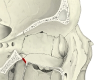 Right orbit, section (lateral view)
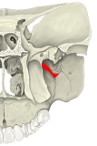 Orbit to palate (coronal section)
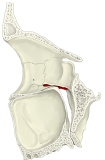 Right orbit, section (medial view)
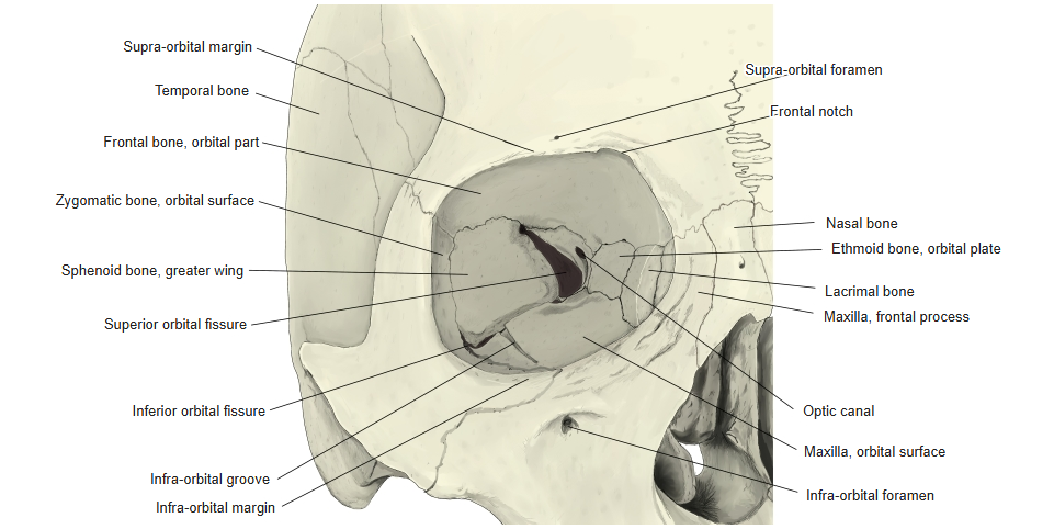
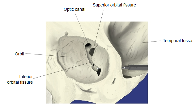
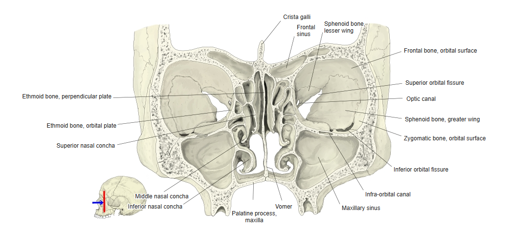
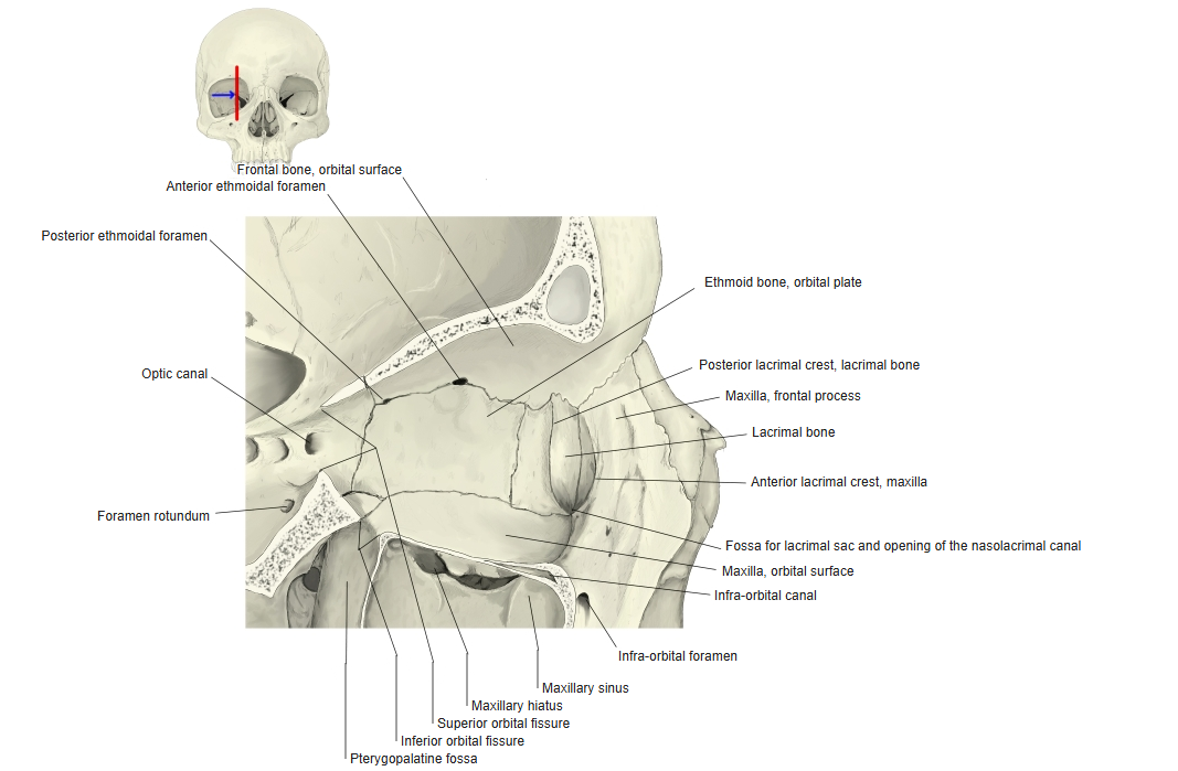
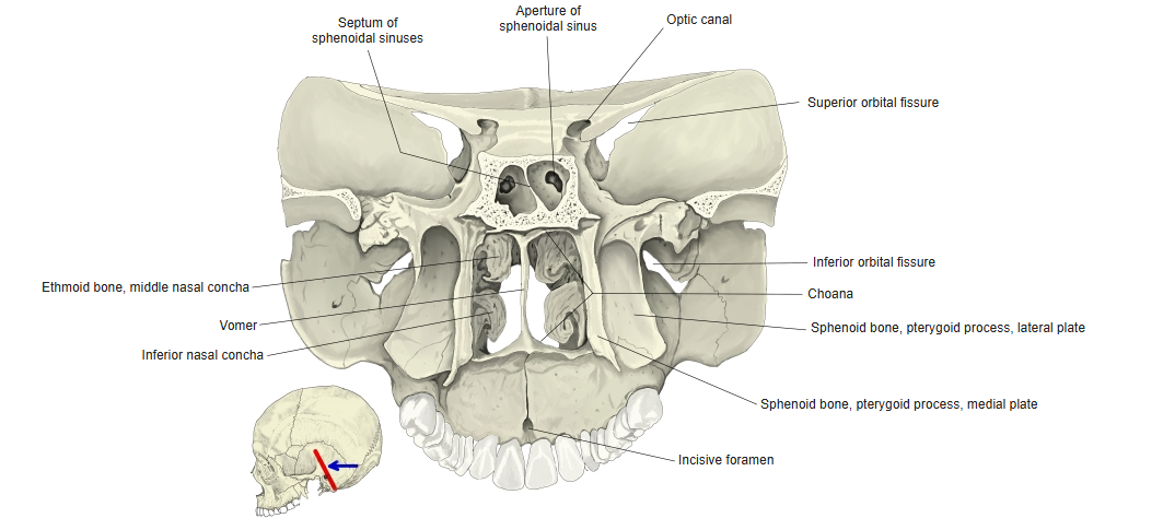
[Structures passing through]
The following nerves and vessels pass through the inferior orbital fissure into the floor of the orbit; the sources consulted differ slightly.
| 1 | Nerves | Zygomatic nerve (branch of the maxillary nerve) | 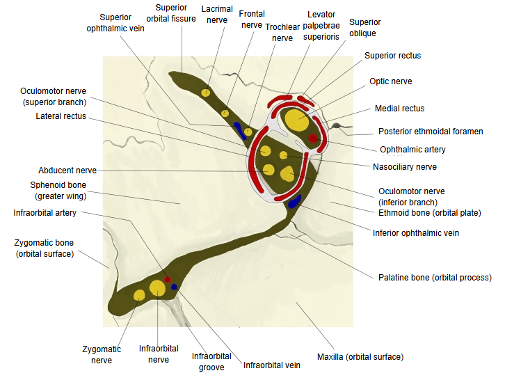 |
| Infra-orbital nerve (continuation of the maxillary nerve) | |||
| 2 | Artery | Infra-orbital artery | |
| 3 | Veins | Infra-orbital vein | |
| · A branch of the inferior ophthalmic vein may pass through it to join the pterygoid venous plexus. · Some sources also list small orbital branches from the pterygopalatine ganglion (via the maxillary nerve) and emissary veins. | |||
Reference websites
[Wikipedia]
· Inferior orbital fissure, Wikipedia.
[Websites with illustrations]
· Human orbit (photograph) (Wikimedia Commons)

· Inferior orbital fissure (Radiopaedia)
· The orbit (Kenhub)
Related terms
- Superior orbital fissure
- Orbit
- Infratemporal fossa
- Infraorbital foramen
- Maxillary nerve
- Infraorbital artery
- Sphenoid bone
References and sources
- Further reading: Inferior orbital fissure, Wikipedia.
- Federative International Programme for Anatomical Terminology (FIPAT). Terminologia Anatomica, 2nd ed. (TA2), 2019.