Glomerular capillary
Overview
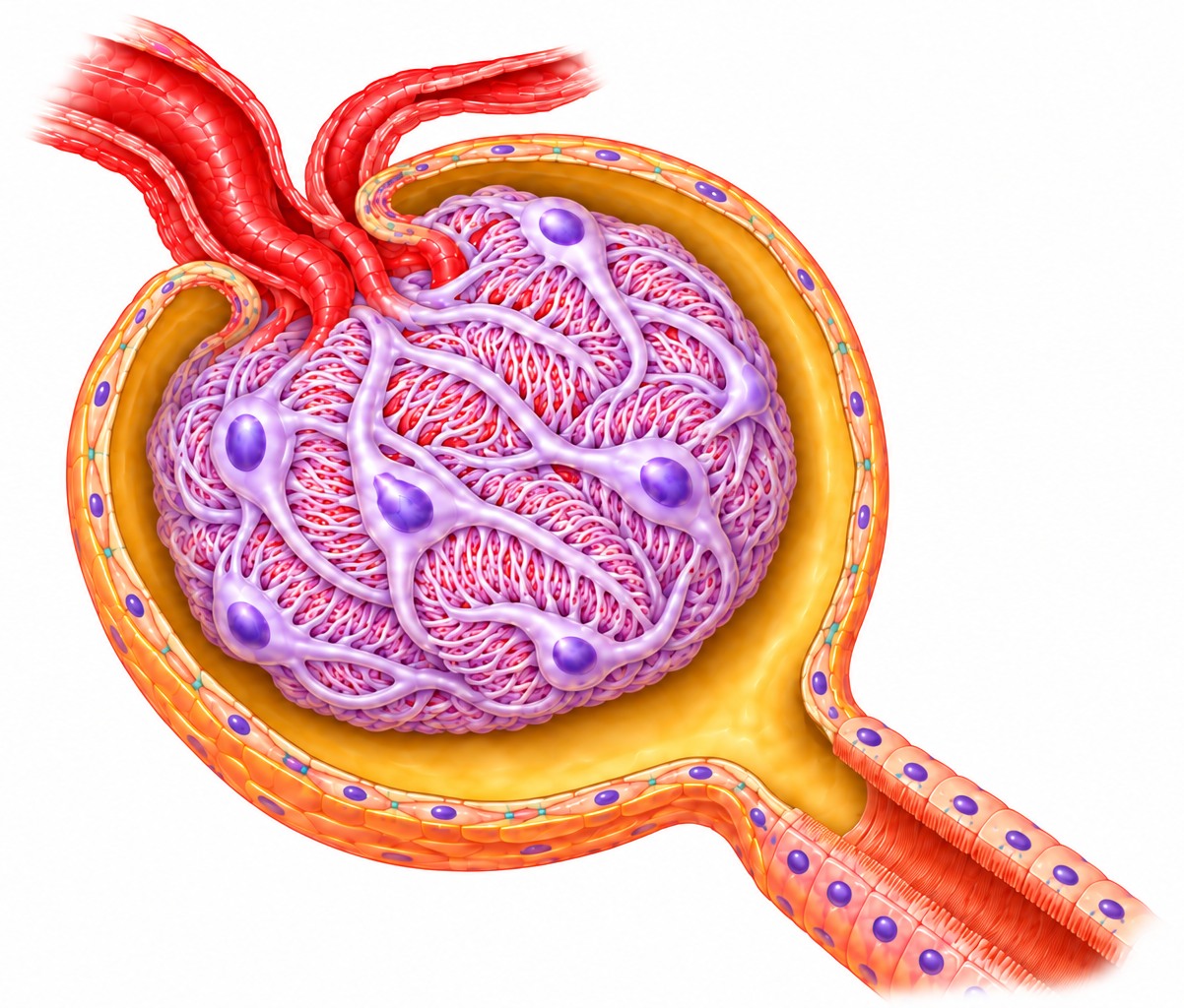
· The glomerular capillaries are the capillaries inside the renal corpuscle that make up the glomerulus: many (several dozen) looping capillaries, grouped into a few lobules and gathered into a tuft. This is where the blood is filtered.
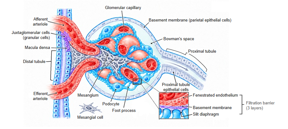
· The afferent arteriole divides into the capillary loops, which join again into a single efferent arteriole that leaves at the vascular pole. They form a special capillary bed between two arterioles and do not drain directly into a vein.
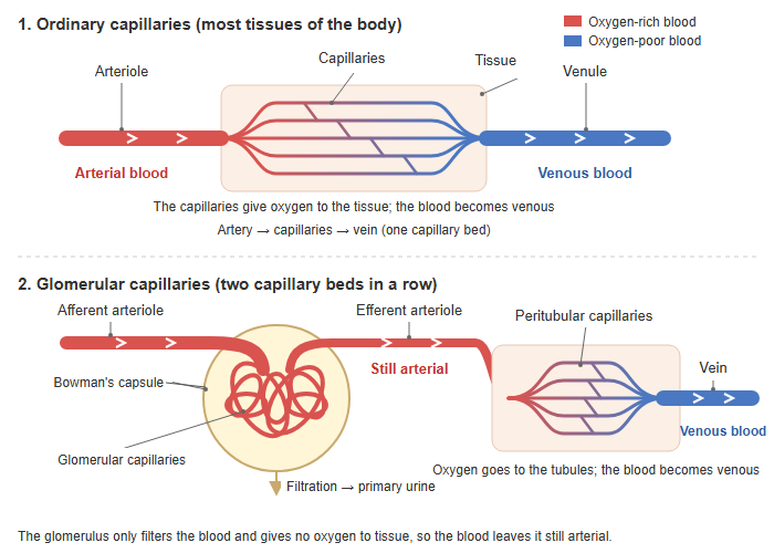
· The resistance of the efferent arteriole keeps the pressure inside the glomerulus high, and this pressure drives filtration.
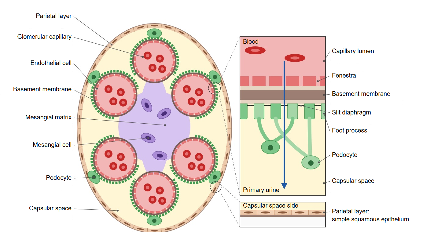
· The wall is made of endothelial cells with pores (fenestrae). Outside it lies the glomerular basement membrane, which is covered in turn by the foot processes of the podocytes. The mesangium supports the capillaries from between the loops.
· Water and small solutes of the blood pass through the endothelial pores, the basement membrane and the gaps between the podocyte foot processes into the capsular space of the glomerular capsule. Blood cells and large proteins cannot pass.
· When the wall of the glomerular capillaries is damaged, protein and blood leak into the urine, as in glomerulonephritis and diabetic nephropathy.
Reference websites
[Wikipedia]
· Glomerulus (kidney), Wikipedia.
· Renal corpuscle, Wikipedia.
[Websites with illustrations]
· Renal corpuscle (diagram) (Wikimedia Commons)

· Glomerulus (histology) (Digital Histology)
· Renal corpuscle (electron micrograph) (Histology Guide)
· The glomerulus (TeachMePhysiology)
Related terms
- Glomerular basement membrane
- Podocyte
- Visceral layer of glomerular capsule
- Parietal layer of glomerular capsule
- Capsular space
- Mesangium
- Vascular pole
- Urinary pole
- Kidney
- Renal artery
- Interlobular arteries of kidney (cortical radiate arteries)
- Renal parenchyma
- Renal veins
- Urinary system
References and sources
- Further reading: Glomerulus (kidney), Wikipedia.
- Further reading: Renal corpuscle, Wikipedia.
- Federative International Programme for Anatomical Terminology (FIPAT). Terminologia Anatomica, 2nd ed. (TA2), 2019.