Pterygoid hamulus (Latin: hamulus pterygoideus)
Also called the hamulus of medial pterygoid plate
- Body : lies in the middle of the sphenoid bone and contains the sphenoidal sinus. It has four surfaces.
- Processes
- Greater wing : the largest process, with three surfaces and three borders; it forms part of the middle cranial fossa and of the side wall of the skull.
- Lesser wing : projects to the left and right from both sides of the front of the body; it forms part of the anterior cranial fossa.
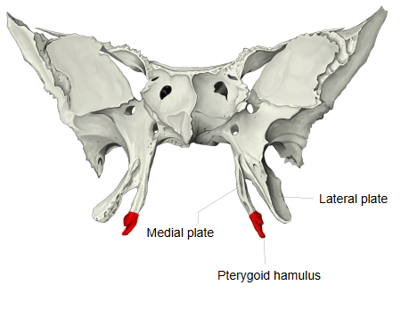
- Pterygoid process : extends downwards from the body and the greater wing; it is made of two plates, medial and lateral.
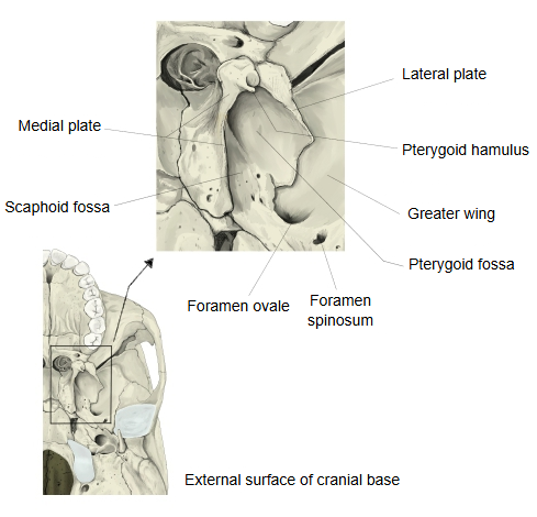
- Medial plate : narrower and longer. Related terms: pterygoid hamulus, groove of pterygoid hamulus, scaphoid fossa, vaginal process, vomerovaginal canal and groove, palatovaginal groove and canal.
- Lateral plate : broader and shorter than the medial plate. Its medial surface gives origin to the deep head of the medial pterygoid muscle and its lateral surface to the lower head of the lateral pterygoid muscle. Related term: pterygospinous process.
- Shared by both plates : pterygoid notch, pterygoid fossa, pterygoid canal.
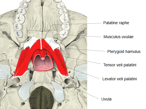
· The groove through which the tendon of tensor veli palatini runs is the groove of the pterygoid hamulus, on the lateral side of the hamulus. After passing through it, the tendon turns sharply medially and spreads into the soft palate as the palatine aponeurosis; the hamulus acts as a pulley.
· It is only a few millimetres long (about 7 mm on average), with a rounded tip; it can be felt through the roof of the mouth behind the last upper molar.
· The levator veli palatini does not arise from the hamulus; it comes from the temporal bone and the auditory tube and passes medial to it.
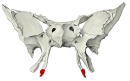 Sphenoid bone (anterior)
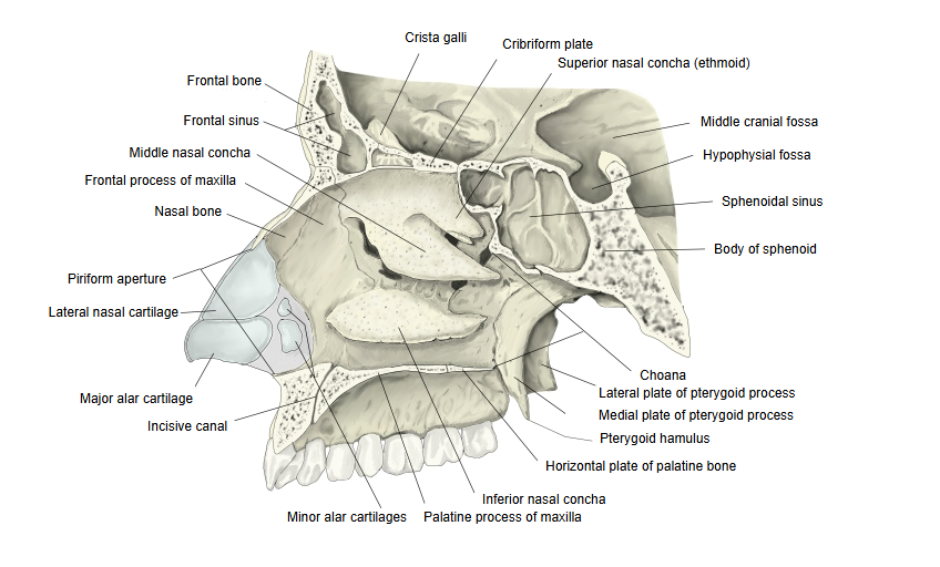
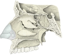 Lateral wall of nasal cavity
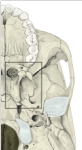 External surface of cranial base
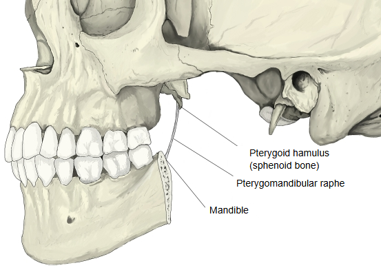
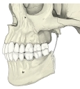 Pterygomandibular raphe
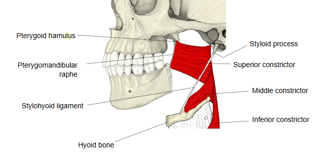
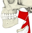 Pharyngeal constrictors
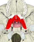 Muscles of the soft palate (inferior)
Reference websites
[Wikipedia]
· Pterygoid hamulus, Wikipedia.
· Pterygoid processes of the sphenoid, Wikipedia.
[Websites with illustrations]
· Sphenoid bone, anterior and inferior surfaces (Gray's Anatomy) (Wikimedia Commons)

· Sphenoid bone, upper and posterior surfaces (Gray's Anatomy) (Wikimedia Commons)

· Base of the skull, outer surface (Gray's Anatomy) (Wikimedia Commons)

Related terms
- Pterygomandibular raphe
- Groove of pterygoid hamulus
- Medial plate of pterygoid process
- Pterygoid process
- Tensor veli palatini
- Palatine aponeurosis
- Soft palate
- Sphenoid bone
- Pharyngeal constrictors
References and sources
- Further reading: Pterygoid hamulus, Wikipedia.
- Federative International Programme for Anatomical Terminology (FIPAT). Terminologia Anatomica, 2nd ed. (TA2), 2019.