Skeletal muscle
- Introduction
- Internal structure
- Shape
- Classification
- Classification by shape
- Classification by action
- Terms related to skeletal muscle
- Lists of skeletal muscles
- Number of muscles in the main regions
- Sensory nerves of skeletal muscle
- Comparison of the muscle types
- Related terms
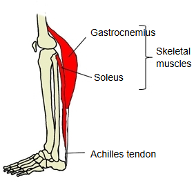
Muscle that is mainly attached to the skeleton and moves it. It is striated and voluntary: it is under conscious control through the motor nerves.
Introduction
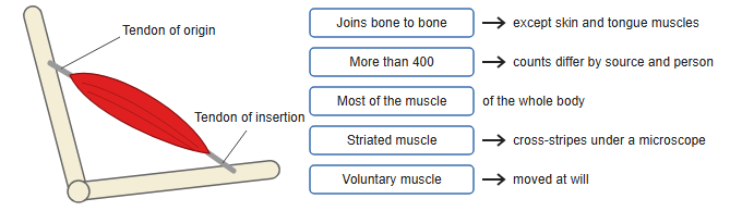
Most skeletal muscles are attached to bones and join one bone to another; they are central to keeping posture and to movement. Some, however, are attached at one end to bone and at the other to another structure, as in the following.
| 1 | Cutaneous (skin) muscles | The muscles of the face, also called the muscles of facial expression, and the platysma of the neck. One end is attached to the skin. |
| 2 | Tongue muscles | The muscles that move the tongue; one end is attached to the tongue itself. |
| 3 | Pharyngeal muscles | The muscles of the wall of the pharynx; they end in the pharyngeal wall and its midline raphe. |
| 4 | Diaphragm | Its fibres end in its own central tendon. |
| 5 | Part of the visceral muscle | Skeletal muscle in the walls of some viscera, such as the upper esophagus. |
Under the microscope its fibres show cross-stripes, because the proteins actin and myosin are laid out in regular repeating units; histologically it is therefore a striated muscle, like cardiac muscle. It makes up most of the body's muscle (about 40% of body weight; some sources give more). It is also a voluntary muscle, controlled at will.
The number of skeletal muscles differs considerably between sources. A standard Japanese anatomy textbook speaks of "about 400 striated muscles", while other sources give much higher numbers. The number of named muscles is often given as about 400 per side or over 600 in total, depending on how muscles are counted.
Internal structure
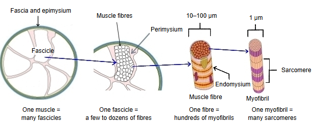
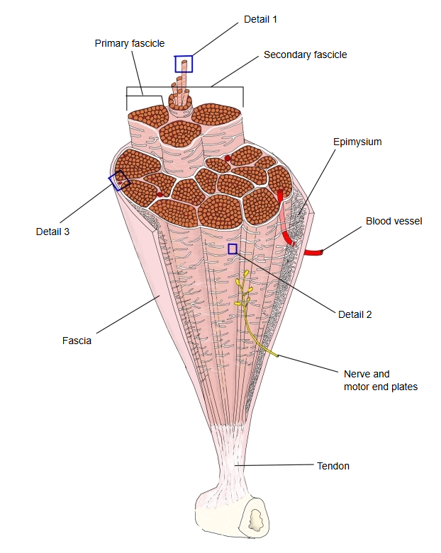
The main levels of structure of a skeletal muscle are as follows.
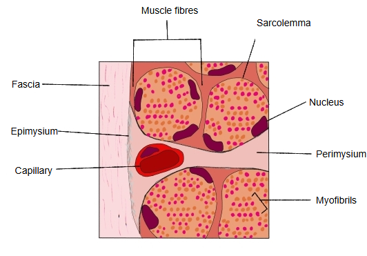
| 1 | Fascicle (muscle bundle) | A skeletal muscle is built of a number of fascicles. They are wrapped together by the epimysium and, outside it, the fascia, forming one muscle with its own name, such as the biceps brachii or the deltoid. See also muscle fascicle. |
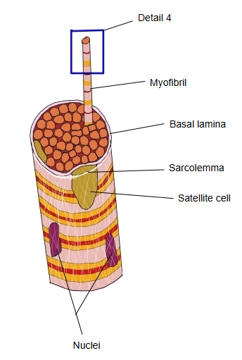
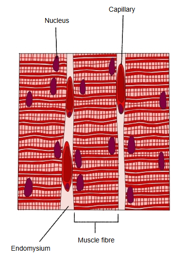
| 2 | Muscle fibre (= muscle cell) | A fascicle is a group of a few to dozens of muscle fibres bound together by the perimysium. Each fibre is one long, multinucleated cell, 10–100 µm thick; its cell membrane is the sarcolemma, and small satellite cells under its basal lamina can repair it. |
| 3 | Myofibril | Each muscle fibre is filled with hundreds to thousands of myofibrils, each about 1 µm thick, and is wrapped by the endomysium. |
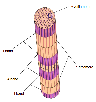
| 4 | Sarcomere | A myofibril is made of many sarcomeres, its repeating contractile units, seen as light I bands and dark A bands. |
Related illustrations:
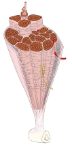 Structure of a skeletal muscle
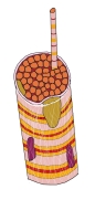 Detail 1: muscle fibre
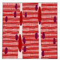 Detail 2: fibres (lengthwise)
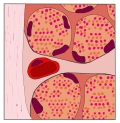 Detail 3: fibres (cross-section)
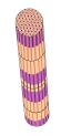 Detail 4: myofibril
Shape
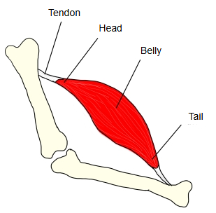
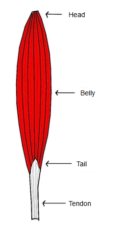
Skeletal muscles have many shapes, but the basic form is the spindle-shaped (fusiform) muscle. The middle part is usually thick and is called the belly; the ends, where the muscle narrows and passes into a tendon, are called the head and the tail.
| 1 | Head | The end nearer the centre of the body, usually the origin. Most muscles have one head, but some have two or more. |
| 2 | Tail | The end opposite the head; it usually inserts on a bone through a tendon. |
| 3 | Belly | The thick, fleshy middle part. Most muscles have one belly, but some have two or more. |
Classification
Skeletal muscles can be classified in two main ways: by their shape and by their action.
- By shape
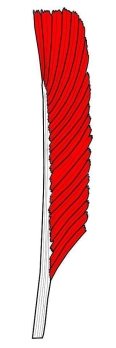
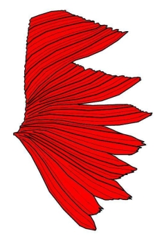
- By form : fusiform, pennate, unipennate, serrate
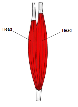
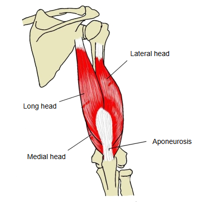
- By number of heads : two-headed, three-headed, four-headed
- By number of bellies : two-bellied, many-bellied
- By number of tails : many-tailed
- By action : flexors, extensors, abductors, adductors, lateral rotators, medial rotators, supinators, pronators
Classification by shape
1. By form
By their form, skeletal muscles can be classified as follows.
| 1 | 2 | 3 | 4 |
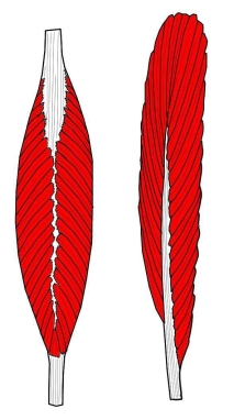
| Fusiform (spindle-shaped) | Pennate (feather-like) | Unipennate (half-feather) | Serrate (saw-toothed) |
| biceps brachii, coracobrachialis, etc. | fibres reach a central tendon from both sides: fibularis longus, etc. | fibres reach the tendon from one side: semimembranosus, etc. | arises by a row of finger-like slips: serratus anterior |
 |  |  |  |
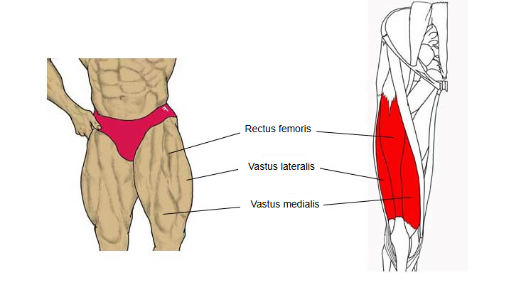
2. By number of heads
By the number of heads, muscles can be classified as follows.
| 1 | 2 | 3 |
| Two-headed muscle (biceps) | Three-headed muscle (triceps) | Four-headed muscle (quadriceps) |
| biceps brachii, etc. | triceps brachii | quadriceps femoris |
 |  |  |
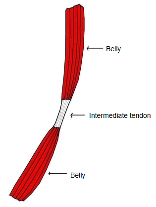
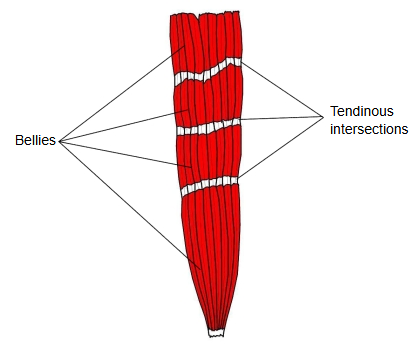
3. By number of bellies
By the number of bellies, muscles can be classified as follows.
| 1 | 2 |
| Two-bellied muscle (digastric): two bellies joined by an intermediate tendon | Many-bellied muscle: several bellies separated by tendinous intersections |
| digastric, omohyoid | rectus abdominis |
 |  |
4. By number of tails
A muscle with several tails (tendons): many-tailed muscle (flexor digitorum profundus, etc.).
Classification by action
Muscles are classified mainly by how they move a joint, as follows.
| 1 | Flexor | A muscle that produces flexion: a movement that decreases the angle of a joint. |
| 2 | Extensor | A muscle that produces extension: a movement that increases the angle of a joint. |
| 3 | Abductor | A muscle that produces abduction: moving the upper or lower limb (lower extremity) away from the trunk. |
| 4 | Adductor | A muscle that produces adduction: moving the upper or lower limb towards the trunk. |
| 5 | Lateral (external) rotator | A muscle that produces lateral rotation. |
| 6 | Medial (internal) rotator | A muscle that produces medial rotation. |
| 7 | Supinator | A muscle that produces supination of the forearm. Seen only in the upper limb. |
| 8 | Pronator | A muscle that produces pronation of the forearm. Seen only in the upper limb. |
| 9 | Others | Sphincters, dilators, levators, depressors, tensors, orbicular (ring) muscles, compressors, etc. |
Terms related to skeletal muscle
Some terms related to skeletal muscle:
| 1 | Cutaneous (skin) muscle | A muscle attached at one end to the skin. In many mammals such muscles are well developed under the skin of the trunk and can move particular areas of skin; in humans they are found only as the muscles of facial expression in the face and head, the platysma in the neck and palmaris brevis in the hand. Some of the facial muscles may be missing. |
| 2 | Agonist | Also called the prime mover: the main muscle that produces a movement. Opposite: antagonist. |
| 3 | Antagonist | A muscle whose action is opposite to that of the muscle producing the movement. |
| 4 | Synergist | A muscle that works together with the agonist to produce the same action. See synergistic muscle. |
| 5 | Biarticular (two-joint) muscle | A muscle whose origin and insertion lie across two joints. |
Lists of skeletal muscles
[List I: all skeletal muscles]
A list of the skeletal muscles with illustrations and links to the page of each muscle: List of skeletal muscles.
1. For each muscle it gives the name, the region and links to an illustration and to the detailed page of the muscle.
[List II: by region]
A standard Japanese anatomy textbook divides the skeletal muscles by position into four large groups:
1. muscles of the head, 2. muscles of the neck, 3. muscles of the trunk, 4. muscles of the limbs.
The muscles of the trunk and of the limbs are then divided further:
· Trunk: muscles of the back, muscles of the thorax, muscles of the abdomen, muscles of the pelvis and muscles of the perineum.
· Limbs: muscles of the upper limb and muscles of the lower limb (lower extremity).
The first volume of that textbook treats the muscles of the pelvis and of the perineum separately, but the second volume puts them together as the muscles of the perineum; this page follows the second volume.
| Region | Number of muscles |
|---|---|
| Head | 29 (+8) |
| Neck | 18 |
| Thorax | 11 |
| Abdomen | 6 |
| Back | 21 |
| Perineum | 10 |
| Upper limb | 41 |
| Lower limb | 54 |
Number of muscles in the main regions
The numbers of muscles in the main regions of the limbs are as follows.
| Upper limb | Number | Lower limb | Number |
|---|---|---|---|
| Shoulder girdle | 6 | Pelvic girdle (hip) | 12 |
| Arm | 5 | Thigh | 16 |
| Forearm | 19 | Leg | 13 |
| Hand | 11 | Foot | 13 |
Sensory nerves of skeletal muscle
Does skeletal muscle have sensory nerves?
Yes, and plenty of them.
Skeletal muscle has not only motor nerves (which make it contract; each motor fibre ends on a muscle fibre at a motor end plate) but also many sensory nerves (which report the state of the muscle to the central nervous system).
Muscle spindles
· Sense how far and how fast the muscle is stretched (its length and the speed of change).
· Mainly group Ia and group II afferent fibres are involved.
· They are the sensors of the stretch reflex (for example the knee jerk, tapped on the patellar tendon).
Golgi tendon organs
· Sense the tension (force) of the muscle.
· Group Ib afferent fibres are involved.
· They take part in a reflex that prevents excessive tension.
Free nerve endings
· Lie in the fascia and within the muscle, and carry pain, temperature and similar sensations.
Skeletal muscle therefore has both motor (efferent) and sensory (afferent) nerves.
The sensory nerves are far from few: they are an essential part of the nerve supply, needed for the control of the muscle.
Comparison of the muscle types
A simple comparison of skeletal muscle, smooth muscle and cardiac muscle:
| Skeletal muscle (striated) | Smooth muscle | Cardiac muscle (striated) | ||
| 1 | Motor end plate | present | absent | absent |
| 2 | Fibres | cylindrical, long (up to about 15 cm) | spindle-shaped, short (up to about 0.2 mm) | branched |
| 3 | Mitochondria | moderate to many (most in red, slow fibres) | few | very many |
| 4 | Nuclei per fibre | many (at the edge of the fibre) | 1 | 1 (sometimes 2) |
| 5 | Sarcomeres | present, length up to 3.65 µm | absent | present, length up to 2.6 µm |
Related terms
- Myofibril
- Muscle
- Striated muscle
- Smooth muscle
- Voluntary muscle
- Muscle fascicle
- Endomysium
- Sarcomere
- List of Skeletal Muscles
References and sources
- Further reading: Skeletal muscle, Wikipedia.
- Further reading: Muscle architecture, Wikipedia.
- Federative International Programme for Anatomical Terminology (FIPAT). Terminologia Anatomica, 2nd ed. (TA2), 2019.