B cell
- Overview
- Before and after activation
- Where B cells are found
- Functions
- Reference websites
- (Wikipedia)
Overview
cytes
- T cells : = T lymphocytes; about 60–70%. Function: cell-mediated immunity (removal of infected cells and tumour cells).
- B cells : = B lymphocytes; about 10–20%. Function: humoral immunity (production of antibodies).
- NK cells : = natural killer cells; about 10–15%. Function: non-specific (innate) immunity, such as the removal of virus-infected cells.
Antigens carried in the blood and lymph include the following.
1. From pathogens: proteins and polysaccharides of bacteria and viruses
2. From cancer cells: tumour-specific and tumour-associated antigens
3. Others: toxins, parasite antigens, vaccine components and others
Life of a B cell
The bone marrow cells come mainly from the pelvis, vertebrae, sternum and ribs; the immature B cells are carried in the blood and lymph to the secondary lymphoid organs (spleen, lymph nodes and others).
· Short-lived plasma cells: most stay where they formed and disappear within a few days to about a week. Long-lived plasma cells: move to the bone marrow and mucosal tissues and produce antibody for months to decades.
· Memory B cells: survive for a long time in a resting state and respond quickly when they meet the same antigen again. Most stay in the secondary lymphoid organs, but some circulate through the body.
· The immunity in which B cells (through the plasma cells they become) act by means of antibodies is called humoral immunity.
cytes
Humoral immunity
Before and after activation
B cells produced in the bone marrow (immature B cells) can produce antibodies only after they have moved to a secondary lymphoid organ and been activated there (activated B cells).
The table compares immature and activated B cells.
| Feature | Immature B cell | Activated B cell | |
|---|---|---|---|
| 1 | Size | Small (6–8 µm) | Medium to fairly large (10–12 µm) |
| 2 | Nucleus | Dense chromatin; nucleolus not prominent | Chromatin dispersed; the nucleolus may be prominent |
| 3 | Cytoplasm | Narrow, pale blue | Wider, strongly basophilic, with well-developed rough endoplasmic reticulum |
| 4 | Function | Resting, still maturing | Activated after recognising the antigen; preparing to differentiate |
| 5 | Appearance | Under the microscope an almost resting lymphocyte | Under the microscope somewhat larger, with a clear nucleus and more cytoplasm |
Where B cells are found
The two main secondary lymphoid organs in which B cells are found are:
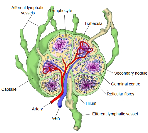
1. Lymph nodes: in the lymphoid nodules of the cortex, where they form germinal centres
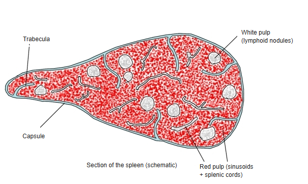
2. Spleen: in the lymphoid nodules of the white pulp, where they form germinal centres
Others: the tonsils, the mucosa-associated lymphoid tissue (for example the Peyer's patches) and the bone marrow (long-lived plasma cells, cells of the B line formed in the secondary lymphoid organs, return to the bone marrow)
 Lymph node
 Section of the spleen
Functions
1. Production of antibodies
B cells cannot attack antigens directly, as T cells and natural killer cells do. Instead, stimulated by cytokines from helper T cells, they become plasma cells, which produce antibodies; the antibodies bind the antigen and so promote the immune response.
2. Antigen presentation
Like dendritic cells and macrophages, B cells are antigen-presenting cells: they break down protein antigens into peptides, bind these to MHC class II molecules and present them on the cell surface, which activates T cells and promotes the immune response.
Antigen presentation (example: a macrophage)
Reference websites
[Wikipedia]
· B cell, Wikipedia.
· Antigen presentation, Wikipedia.