Renal corpuscle (Latin: corpusculum renale)
Also called the Malpighian corpuscle (Malpighian body)
- Overview
- Components
- Location
- The two poles
- How the primary urine is made
- Reference websites
- (Wikipedia, websites with illustrations)
- Related terms
Overview
· The renal corpuscle and the renal tubule that follows it together form a nephron.
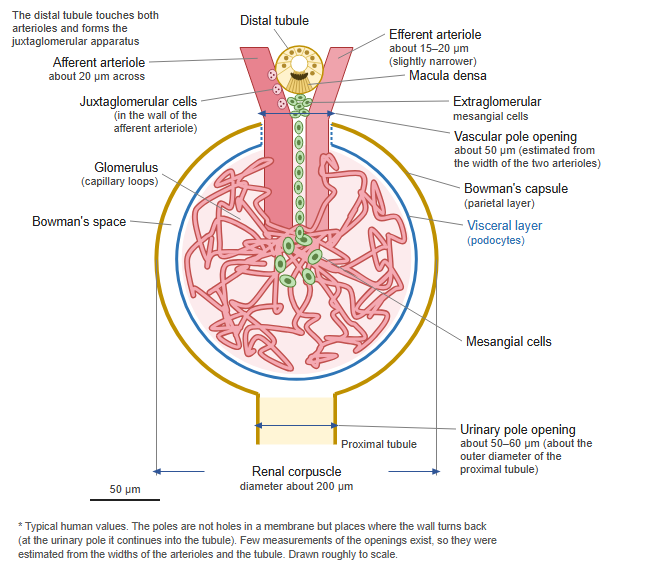
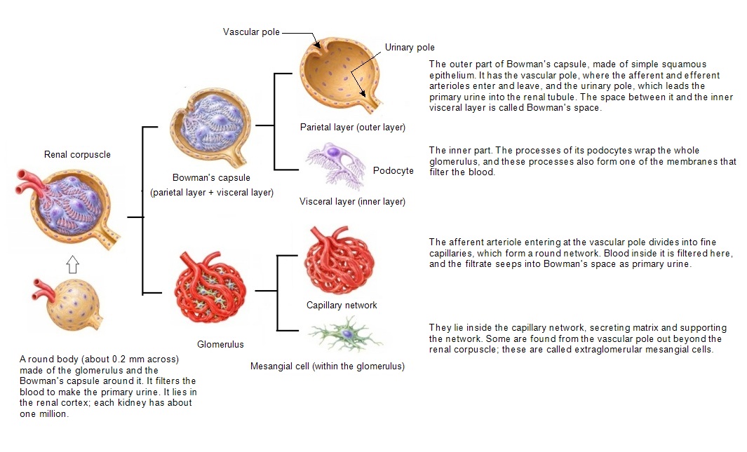
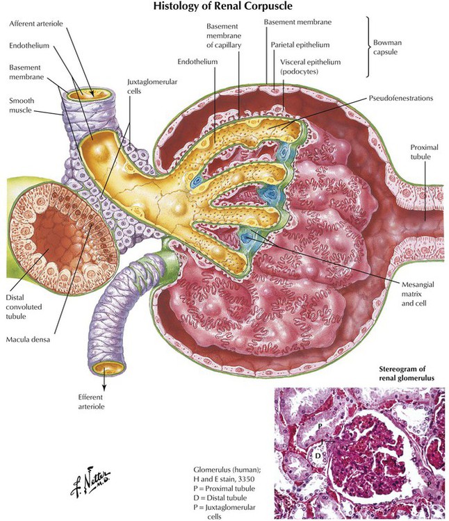
· Fluid filtered in the glomerulus collects in the cavity of Bowman's capsule (the capsular space, or Bowman's space) and flows from the urinary pole into the renal tubule.
· Near the vascular pole, the macula densa of the distal tubule and the juxtaglomerular cells of the afferent arteriole, with other cells, form the juxtaglomerular apparatus, which helps to regulate blood pressure and glomerular filtration.
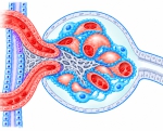 Renal corpuscle (section)
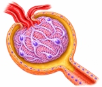 Renal corpuscle (section)
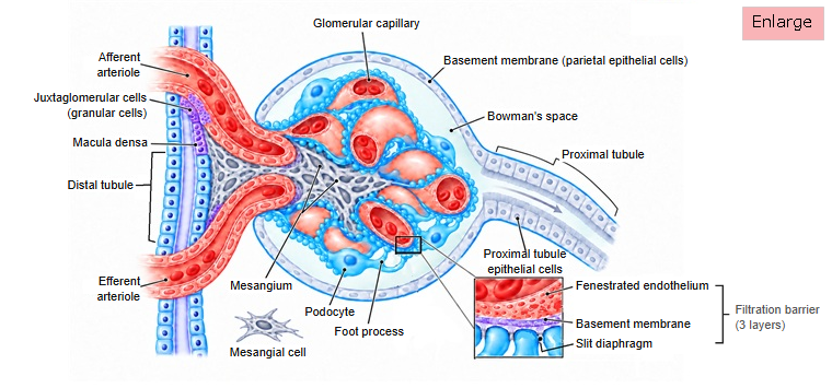
Components
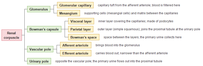
The chart below shows the whole renal corpuscle in a simple form.
corpuscle
- Glomerulus ( Glomerular capillary Mesangium )
- Bowman's capsule ( Visceral layer Parietal layer Bowman's space )
- Vascular pole ( Afferent arteriole Efferent arteriole )
- Urinary pole Detailed chart Whole kidney
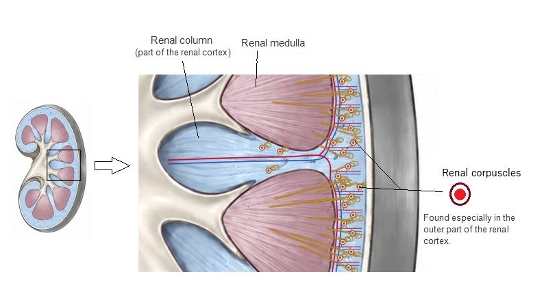
Location
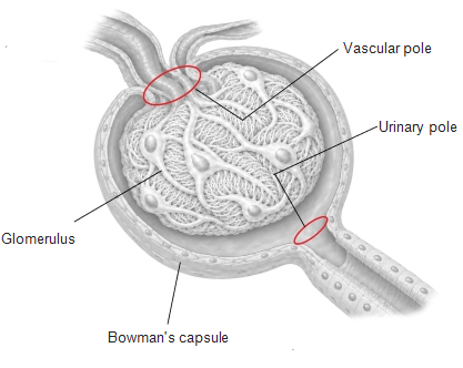
The two poles
| 1 | Vascular pole | The afferent arteriole enters and the efferent arteriole leaves (the way in and out for the blood of the glomerulus). |
| 2 | Urinary pole | Opposite the vascular pole. The proximal tubule begins here, and the primary urine in Bowman's space flows out this way. |
How the primary urine is made
An adult man has only about 4.5–5 L of blood in all, but far more blood than that passes through the kidneys each day.
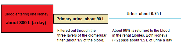
Blood that enters the glomerulus under high pressure is filtered into the primary urine as follows.
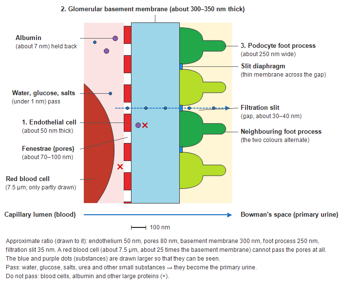
1. Pushed by the blood pressure in the glomerular capillaries (about 50–60 mmHg), water and small substances of the plasma (glucose, amino acids, salts, urea and others) pass through the three layers of the filter (1: the pores of the endothelial cells, 2: the glomerular basement membrane, 3: the slit diaphragms of the podocytes) into Bowman's space. This is the primary urine.
2. Blood cells such as red blood cells, and large proteins such as albumin, cannot pass the filter; they stay in the blood vessels and leave through the efferent arteriole.
3. The primary urine collected in Bowman's space flows out from the urinary pole into the proximal tubule.
Glomerular filtration
Pass: water, glucose, salts and other small substances. Do not pass: blood cells and large proteins.
Note: both kidneys together receive about 1,500–1,700 L of blood a day (about 20–25% of the blood pumped by the heart). The filtrate seeps out over the whole surface of the glomerulus; about 80% of the plasma is not filtered and leaves through the efferent arteriole.
Note: the blood leaves the glomerulus not through a vein but through the narrow efferent arteriole (a capillary bed between two arterioles, called an arterial rete mirabile; in humans found only in the glomerulus), so the pressure in the glomerulus stays about 2–3 times that of ordinary capillaries and filtration goes on.
Note: about 150–180 L of primary urine is made a day, but about 99% of it is reabsorbed in the renal tubules; only about 1.5 L becomes urine.
Reference websites
[Wikipedia]
· Renal corpuscle, Wikipedia.
· Nephron, Wikipedia.
[Websites with illustrations]
· Glomerular filtration (University of Chicago Kidney Stone Program)
· Renal corpuscle (2D functional tissue unit) (Human Reference Atlas)
· Renal corpuscle (stock illustration) (Alamy)
· Renal corpuscle (textbook figure) (Basicmedical Key)
Related terms
- Glomerulus
- Glomerular capsule
- Capsular space
- Vascular pole
- Urinary pole
- Juxtaglomerular apparatus
- Nephron
References and sources
- Further reading: Renal corpuscle, Wikipedia.
- Further reading: Nephron, Wikipedia.
- Federative International Programme for Anatomical Terminology (FIPAT). Terminologia Anatomica, 2nd ed. (TA2), 2019.