Muscles of auditory ossicles (Latin: musculi ossiculorum auditus)
Also called the muscles of the middle ear
of the head
- Facial muscles
- Muscles of mastication : four
- Extra-ocular muscles
- Muscles of the auditory ossicles : two – stapedius and tensor tympani
- Muscles of the tongue
- Muscles of the soft palate and fauces
See also: List of the muscles of the head.
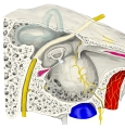 Right ear, lateral view
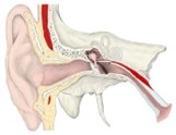 Ear (coronal section)
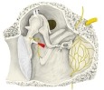 Walls of the tympanic cavity
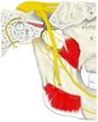 Mandibular nerve, medial view
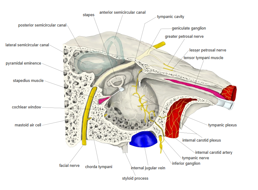
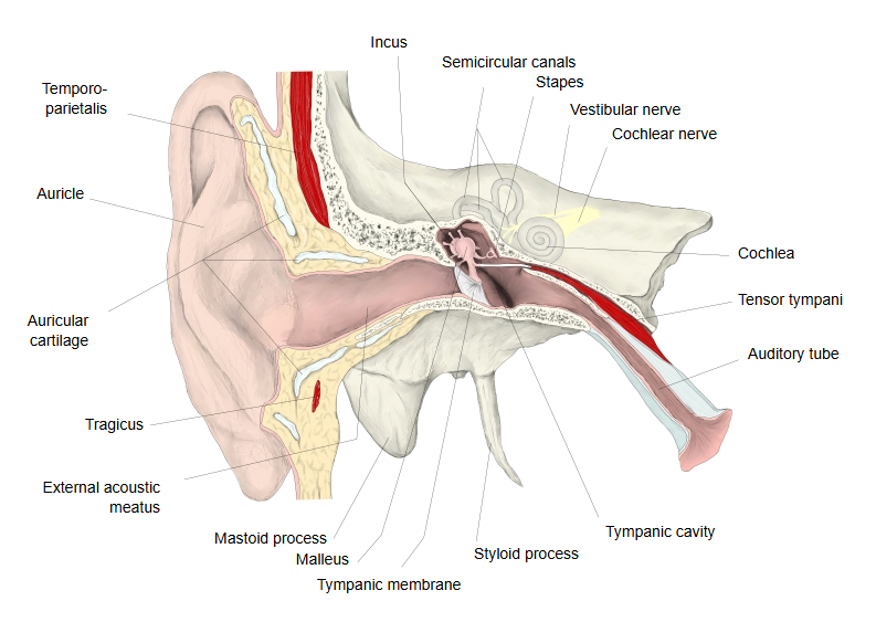
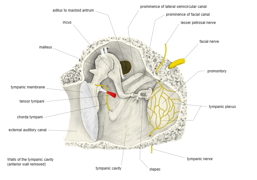
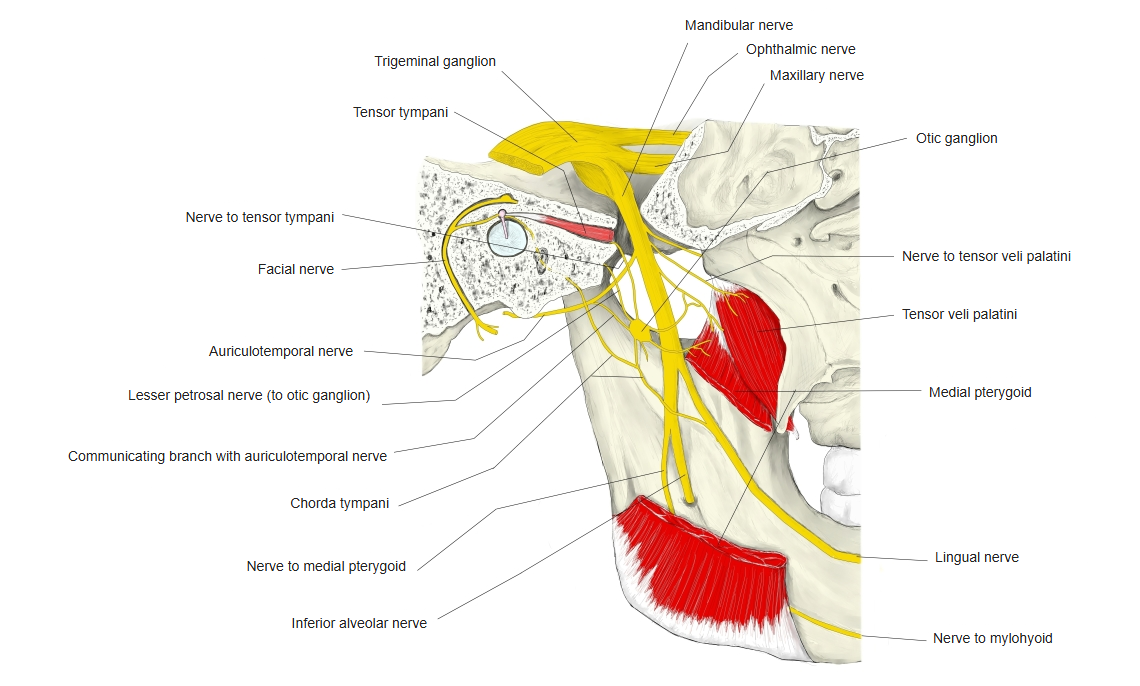
The muscles of the auditory ossicles attach to the auditory ossicles and move them; they are the tensor tympani and the stapedius. The tensor tympani arises from the walls of the canal for tensor tympani in the temporal bone, enters the tympanic cavity and inserts on the malleus; it pulls the handle of the malleus medially and so tenses the tympanic membrane. It is supplied by the mandibular nerve. The stapedius arises inside the pyramidal eminence of the tympanic cavity and inserts on the neck of the stapes. It is supplied by the nerve to stapedius, a branch of the facial nerve. Both damp the vibration of the ossicles; the stapedius contracts in the acoustic reflex and limits the transmission of loud sounds to the internal ear.
Related terms
- Auditory ossicles
- Middle ear
- Tympanic membrane
- Pyramidal eminence
- Canal for tensor tympani
- Tensor tympani
- Stapedius
References and sources
- Further reading: Tensor tympani muscle, Wikipedia.
- Further reading: Stapedius muscle, Wikipedia.
- Federative International Programme for Anatomical Terminology (FIPAT). Terminologia Anatomica, 2nd ed. (TA2), 2019.