Latin: ulna · one of the two bones of the forearm
Overview
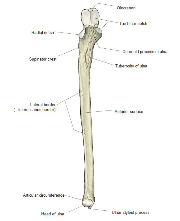
- A long bone on the medial (little-finger) side of the forearm, parallel to the radius (about 24 cm in men, 21–22 cm in women).
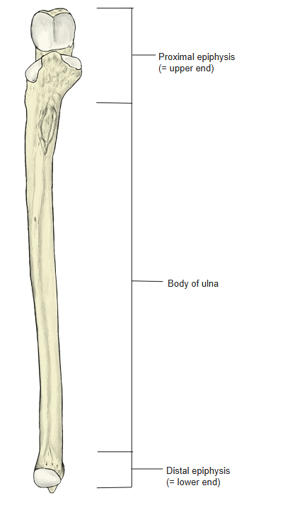
- It has an upper end, a shaft (body of ulna) and a lower end. Unlike the radius, the upper end is large and the lower end is slender.
- The upper end bears the olecranon and coronoid process, which together form the trochlear notch for the humerus.
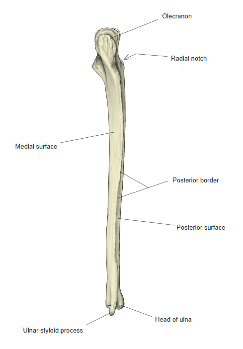
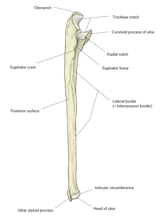
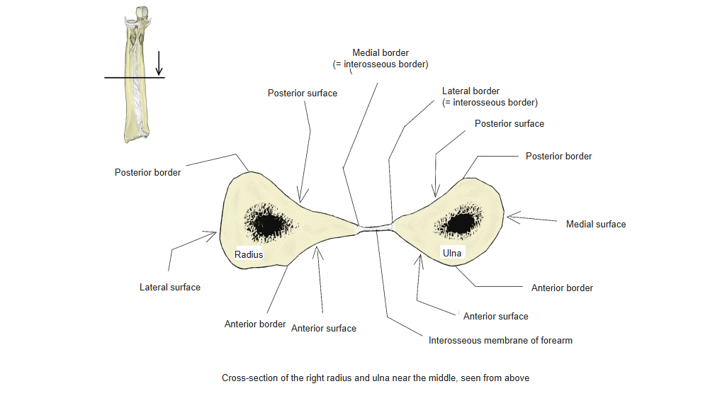
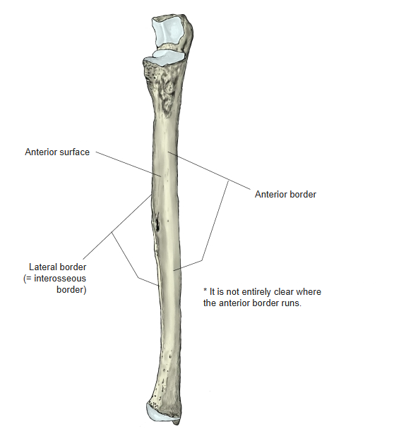
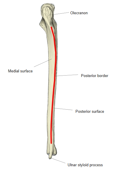
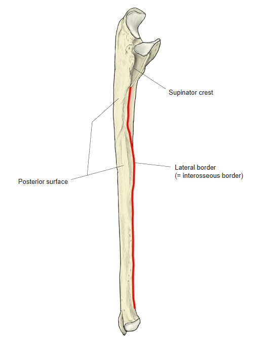
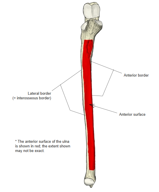
- The shaft has three borders and three surfaces; the lateral border is sharp and is called the interosseous border.
- Anomaly: part or all of the ulna may be absent, and it may be fused with the radius.
Right ulna 
Upper end of the ulna
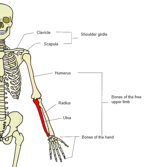
- Bones of the upper limb (one side)
- Shoulder girdle
- Clavicle
- Scapula
- Free upper limb
- Arm
- Humerus
- Forearm
- Radius ← thumb side
- Ulna ← little-finger side
- Hand
- Carpal bones (8)
- Metacarpal bones (5)
- Phalanges (14)
- Arm
- Shoulder girdle
Illustrations

Right ulna (anterior) 
Right ulna (posterior) 
Ulna (lateral) 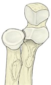
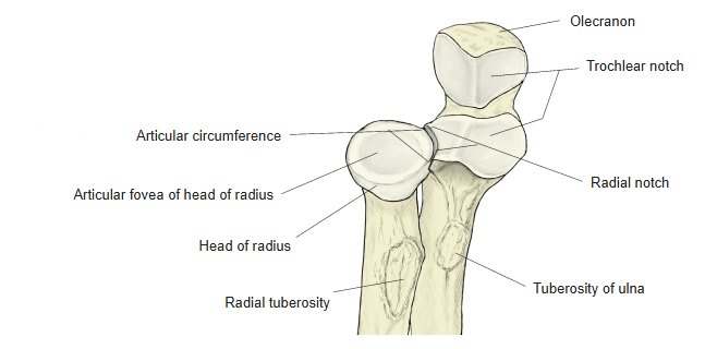
Radius and ulna (proximal ends) 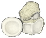
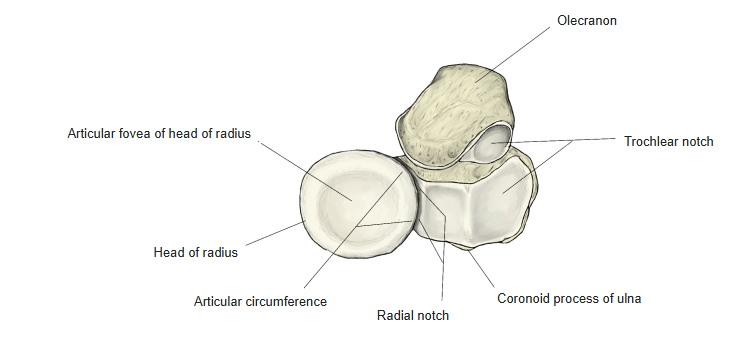
Radius and ulna, seen from above 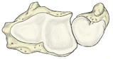
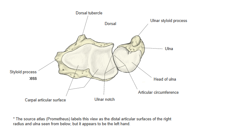
Radius and ulna, seen from below 
Cross-section of the forearm bones
Articulations
| # | Bone | Joint |
|---|---|---|
| 1 | Humerus | Humeroulnar joint |
| 2 | Radius | Proximal radioulnar joint |
| 3 | Radius | Distal radioulnar joint |
Elbow joint
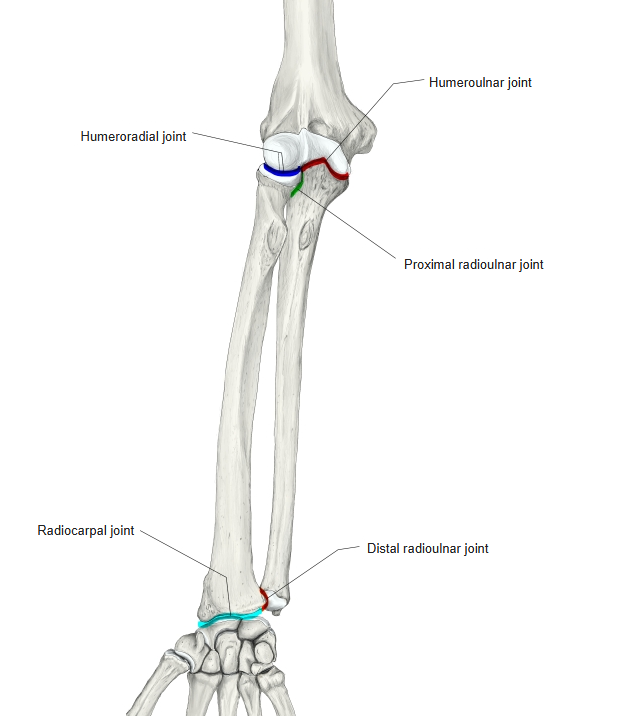
Humeroulnar joint: trochlear notch ↔ trochlea of the humerus. Proximal radioulnar joint: radial notch ↔ articular circumference of the radius. Distal radioulnar joint: articular circumference of the head ↔ ulnar notch of the radius.
Ligaments
The following ligaments attach to the ulna. The list is intended to be comprehensive but may omit minor ligaments.
| # | Ligament | Connects |
|---|---|---|
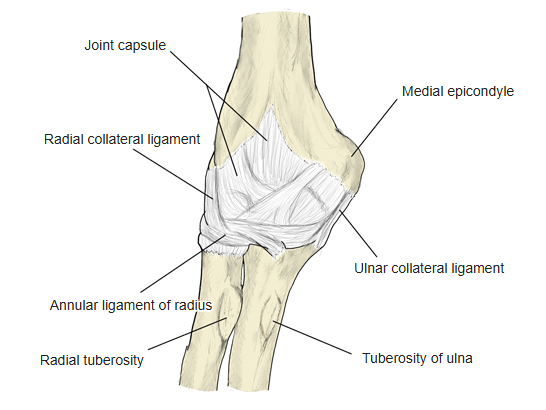
| 1 | Ulnar collateral ligament of the elbow (anterior, posterior and transverse parts) | Coronoid process, olecranon – medial epicondyle of humerus |
| 2 | Radial collateral ligament of the elbow | Lateral epicondyle of humerus – annular ligament of radius (some fibres reach the lateral margin of the ulna) |
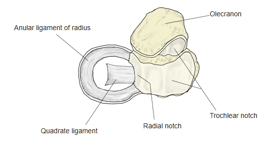
| 3 | Anular ligament of radius | Joins the anterior and posterior margins of the radial notch |
| 4 | Quadrate ligament | Radial notch – neck of radius |
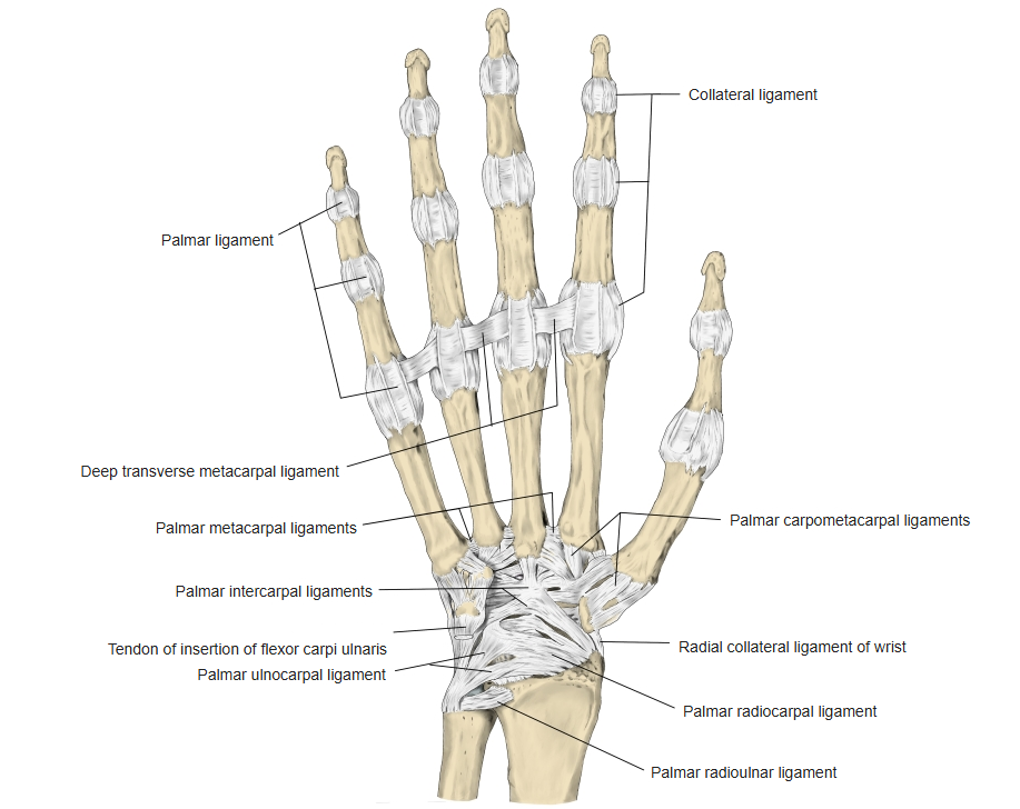
| 5 | Palmar radioulnar ligament | Head of ulna – ulnar notch of radius |
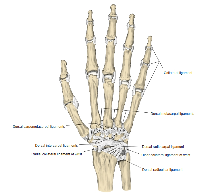
| 6 | Dorsal radioulnar ligament | Styloid process of ulna – medial side of the lower end of radius |
| 7 | Ulnar collateral ligament of the wrist | Styloid process of ulna – triquetrum |
| 8 | Palmar ulnocarpal ligament | Styloid process of ulna – triquetrum, capitate, pisiform |
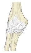
Right elbow joint (posterior) 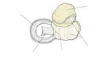
Quadrate ligament 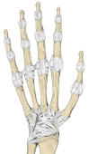
Right hand (palm) 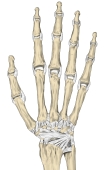
Right hand (dorsum)
Muscle attachments
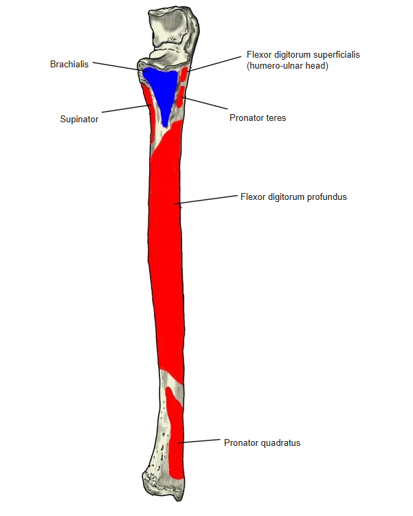
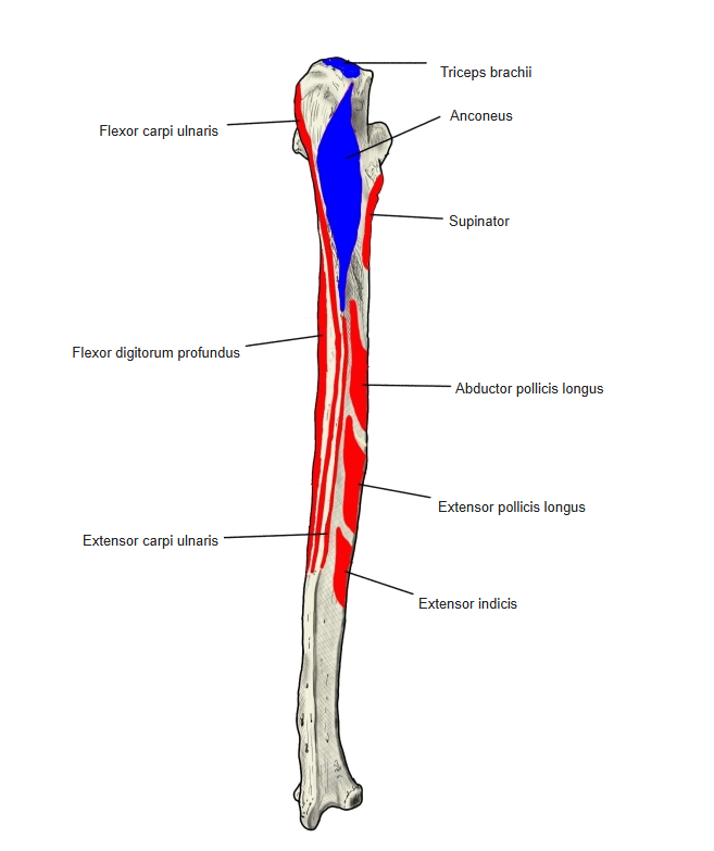
- Origin of: Pronator teres, Supinator, Pronator quadratus, Flexor carpi ulnaris, Extensor carpi ulnaris, Abductor pollicis longus, Flexor digitorum superficialis, Flexor digitorum profundus, Flexor pollicis longus, Extensor pollicis longus, Extensor indicis
- Insertion of: Brachialis, Triceps brachii, Anconeus
Origins: pronator teres (ulnar head) — coronoid process; supinator — supinator crest; pronator quadratus — anterior surface, lower quarter; flexor carpi ulnaris (ulnar head) — medial margin of the olecranon and upper two-thirds of the posterior border; extensor carpi ulnaris (ulnar head) — posterior border (by an aponeurosis); abductor pollicis longus — posterior surface (lateral part) and the interosseous membrane; flexor digitorum superficialis (humeroulnar head) — medial side of the coronoid process; flexor digitorum profundus — anterior and medial surfaces, upper three-quarters; flexor pollicis longus (variable accessory head) — coronoid process; extensor pollicis longus — posterior surface, middle; extensor indicis — posterior surface, below the middle. Insertions: brachialis — coronoid process and tuberosity of ulna; triceps brachii and anconeus — olecranon.

Muscle attachments (anterior) 
Muscle attachments (posterior)
Borders
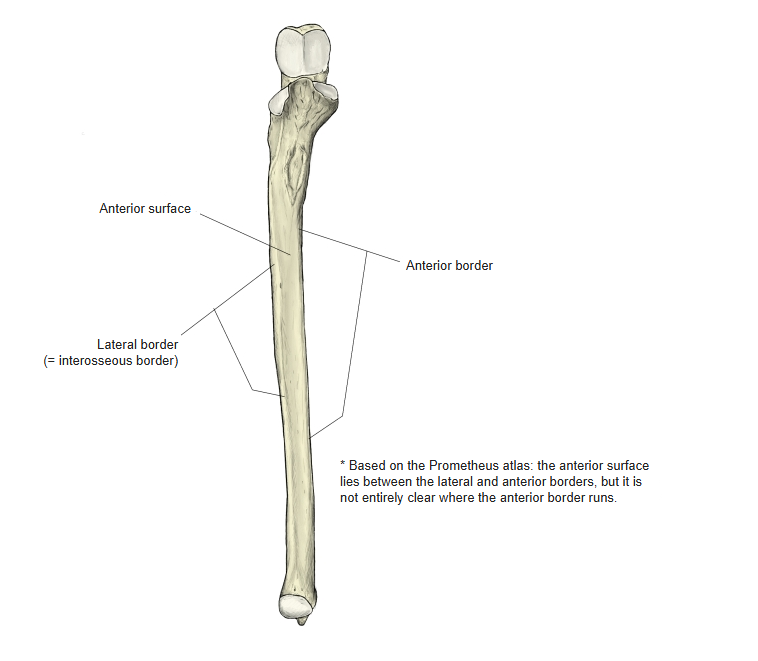
- Anterior border
- Posterior border
- Interosseous (lateral) border — sharp; gives attachment to the interosseous membrane.

Anterior border 
Anterior border (anterior view) 
Posterior border 
Interosseous border
Surfaces
- Anterior surface
- Posterior surface
- Medial surface
Anterior surface
References and sources
- Further reading: Ulna, Wikipedia.