Latin: costae
Overview
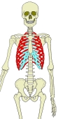
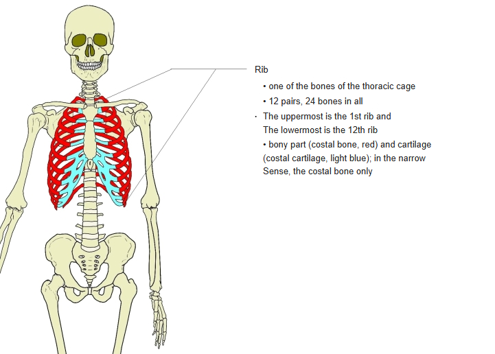
- Flat, curved bones of the thoracic cage (some texts class them as long bones). The term "rib" strictly covers the bony part (costal bone) and the costal cartilage, but in everyday use it usually means the bony part.
- There are 12 pairs, matching the number of thoracic vertebrae.
- Ribs 3–9 have the typical form; ribs 1, 2, 10, 11 and 12 are atypical.
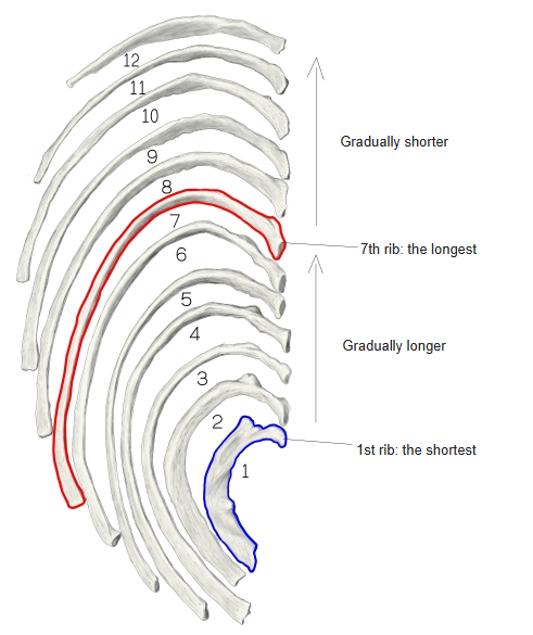
- Length increases from the 1st rib (the shortest) to the 7th (the longest), then decreases again from the 8th.
- Each rib runs downwards and forwards from the spine, then turns upwards near the sternum; in ribs 1 and 2 the bend is at the junction with the costal cartilage, while in the others it is within the cartilage.
The ribs 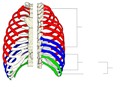
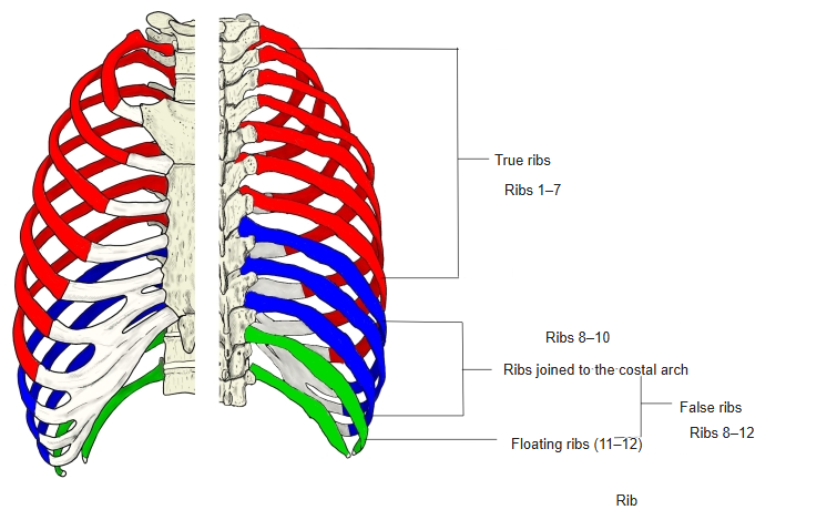
True, false and floating ribs 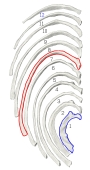
Length of the ribs
- Bones of the trunk
- Vertebral column
- Cervical vertebrae (7)
- Thoracic vertebrae (12)
- Lumbar vertebrae (5)
- Sacrum (1)
- Coccyx (1)
- Thoracic cage
- Sternum (1)
- Ribs (24)
- Vertebral column
Illustrations
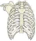
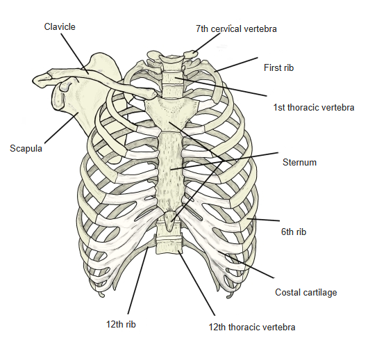
Thorax (anterior) 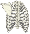
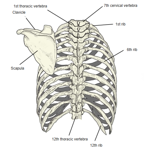
Thorax (posterior) 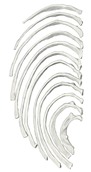
Right ribs (from above) 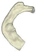
Right 1st rib (from above) 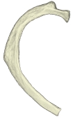
Right 2nd rib (from above) 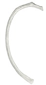
Right 5th rib (from above) 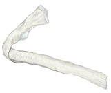
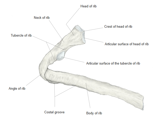
A typical rib (from behind) 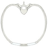
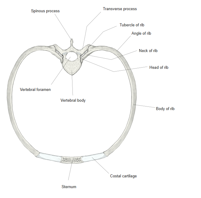
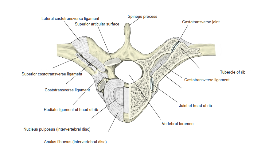
6th rib and thoracic vertebra (from above)
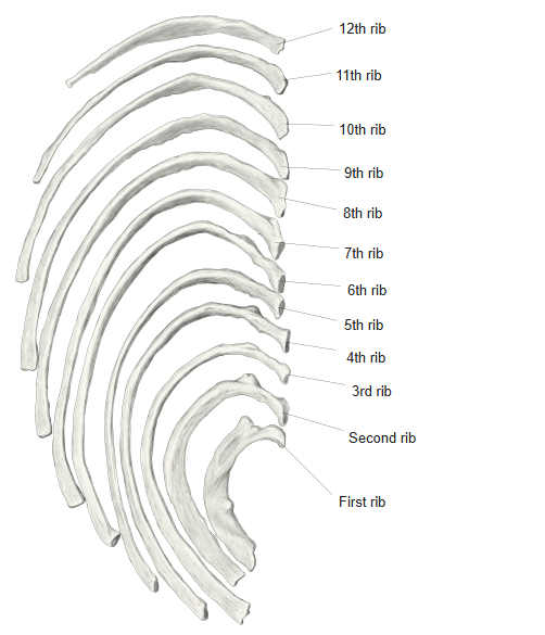
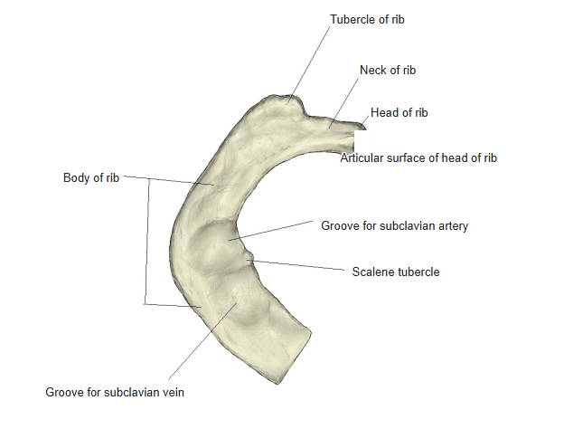
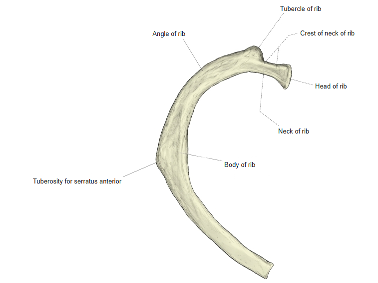
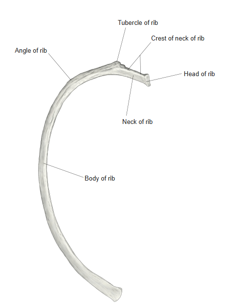
Articulations
| # | Bone | Joint |
|---|---|---|
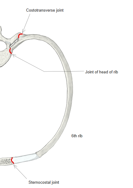
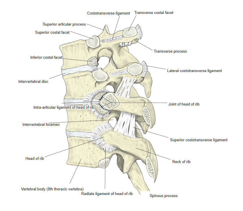
| 1 | Head of rib ⇔ vertebral bodies (costal facets) | Joint of head of rib |
| 2 | Tubercle of rib ⇔ transverse process (transverse costal facet) | Costotransverse joint |
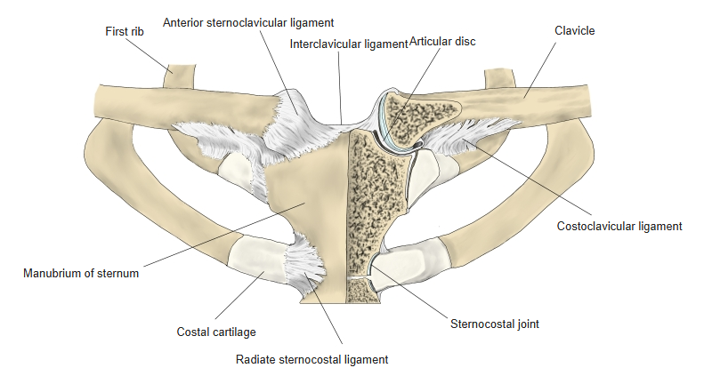
| 3 | Costal cartilage ⇔ sternum (costal notch) | Sternocostal joint |
Joints of the ribs
Ligaments
The following ligaments attach to the ribs. The list is intended to be comprehensive but may omit minor ligaments.
| # | Ligament | Connects |
|---|---|---|
| 1 | Radiate ligament of head of rib | Head of rib – vertebral bodies and intervertebral disc |
| 2 | Intra-articular ligament of head of rib | Crest of head of ribs 2–10 – intervertebral disc |
| 3 | Costotransverse ligament | Neck of rib – transverse process |
| 4 | Superior costotransverse ligament | Neck of rib – transverse process of the vertebra above |
| 5 | Lateral costotransverse ligament | Tubercle of rib – transverse process |
| 6 | Costoclavicular ligament | 1st rib (upper border) – clavicle |
| 7 | Lumbocostal ligament | 12th rib – costal processes of L1 and L2 |
| 8 | Radiate sternocostal ligaments | Costal cartilages – sternum |
| 9 | Intra-articular sternocostal ligament | Costal cartilage – costal notch of the sternum |
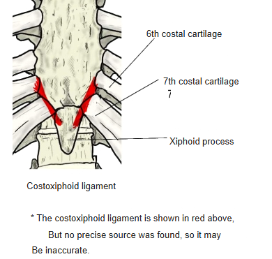
| 10 | Costoxiphoid ligaments | 6th and 7th costal cartilages – xiphoid process |
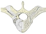
Costovertebral ligaments (from above) 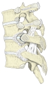
Costovertebral ligaments (lateral) 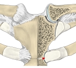
Sternoclavicular and sternocostal ligaments 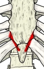
Costoxiphoid ligaments
Muscle attachments
- Origin of: Internal intercostal, External intercostal, Innermost intercostal, Serratus anterior, Subclavius, Pectoralis major, External oblique, Latissimus dorsi, Transversus abdominis, Diaphragm, Subcostales, Transversus thoracis
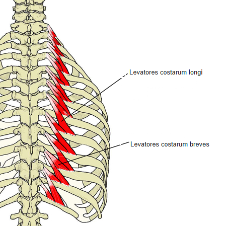
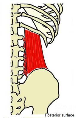
- Insertion of: Internal intercostal, External intercostal, Innermost intercostal, Scalenus anterior, Scalenus medius, Scalenus posterior, Levatores costarum, Serratus posterior superior, Serratus posterior inferior, Iliocostalis, Quadratus lumborum, Rectus abdominis, Subcostales
The intercostal muscles run between neighbouring ribs, so they both arise from and insert on ribs. Scalenus anterior and medius insert on the 1st rib, scalenus posterior on the 2nd, quadratus lumborum on the 12th; rectus abdominis inserts on costal cartilages 5–7. Pectoralis major, transversus abdominis, transversus thoracis and the diaphragm arise partly from the costal cartilages.
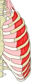
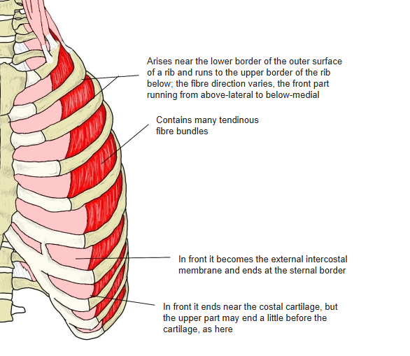
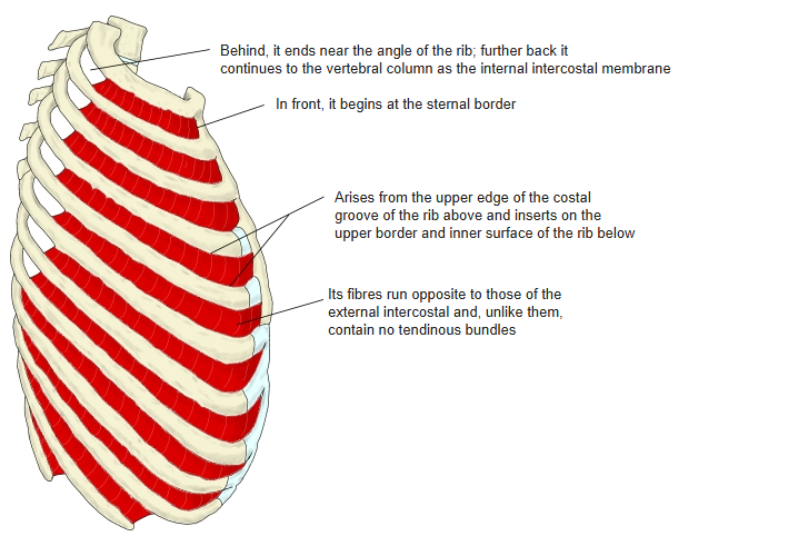
External intercostal 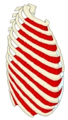
Internal intercostal 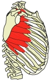
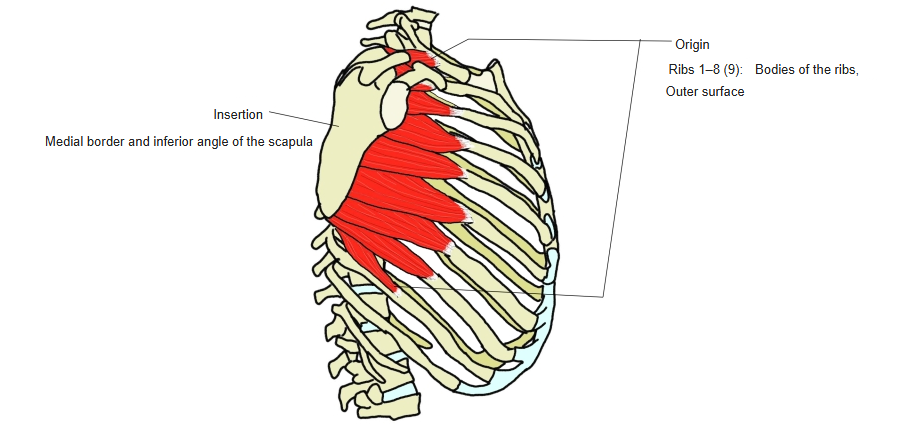
Serratus anterior 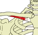
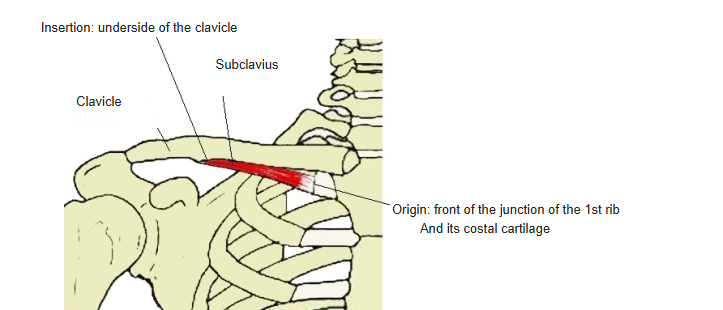
Subclavius 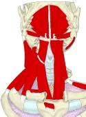
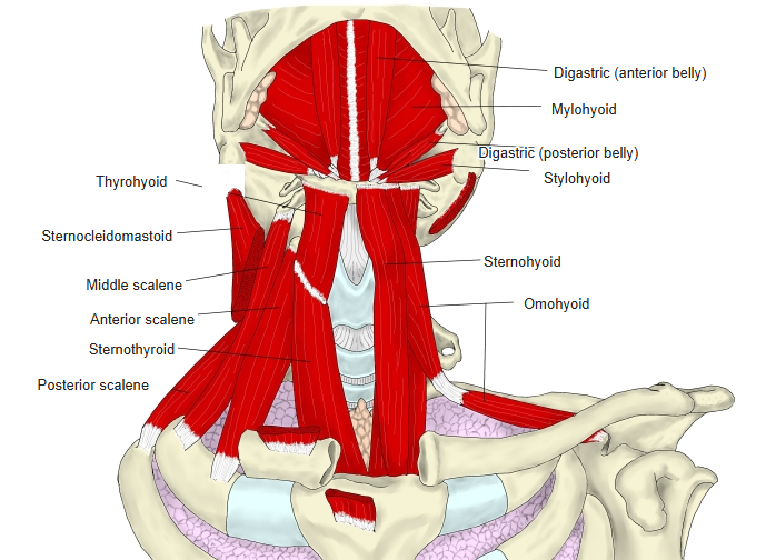
Muscles of the neck (anterior) 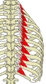
Levatores costarum 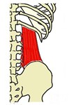
Quadratus lumborum
True, false and floating ribs
- True ribs (1–7) – reach the sternum through their own costal cartilages.
- False ribs (8–12) – do not reach the sternum directly; ribs 8–10 join the cartilage above and form the costal arch.
- Floating ribs (11–12) – have no anterior attachment; their tips end among the muscles of the abdominal wall.
Features of individual ribs
- 1st rib – the shortest, broadest and most strongly curved; it has the groove for the subclavian vein, the scalene tubercle and the groove for the subclavian artery, but no costal groove, and no angle separate from the tubercle.
- 2nd rib – the tubercle and the angle are at the same place; it has the tuberosity for serratus anterior.
- 3rd–9th ribs – typical ribs: head, neck, tubercle, body, angle and costal groove; the 7th is the longest.
- 10th rib – like a typical rib, but its head has only one articular facet.
- 11th and 12th ribs – short, with no neck or tubercle; the 12th may have no angle or costal groove.
Parts of a typical rib
- Head – with an articular surface (divided by the crest of the head) for the bodies of two vertebrae.
- Neck – the narrow part beyond the head, with the crest of the neck.
- Tubercle – on the back, with an articular surface for the transverse process.
- Body (shaft) – with the angle where it turns forwards, and the costal groove on the inner surface of its lower border for the intercostal vessels and nerve.
References and sources
- Further reading: Rib, Wikipedia.