Bare area (Latin: area nuda hepatis)
Also called the bare area of liver
· Outside the bare area, the diaphragm and the liver are both covered by smooth peritoneum, so they can slide freely against each other.
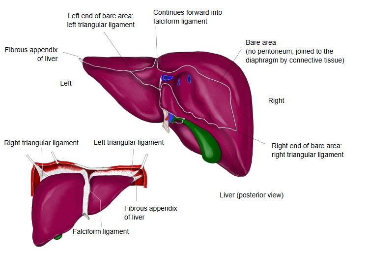
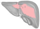 Liver (posterior view)
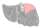 Liver (inferior view)
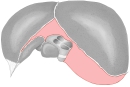 Liver (superior view)
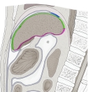 Liver (sagittal section)
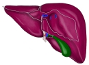 Liver (posterior view)
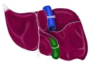 Liver (visceral surface)
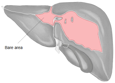
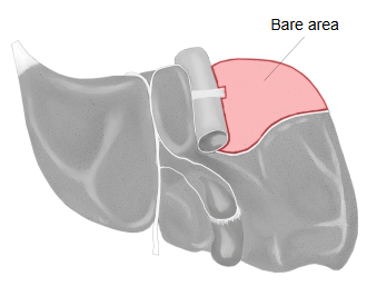
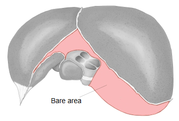
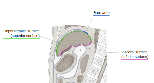
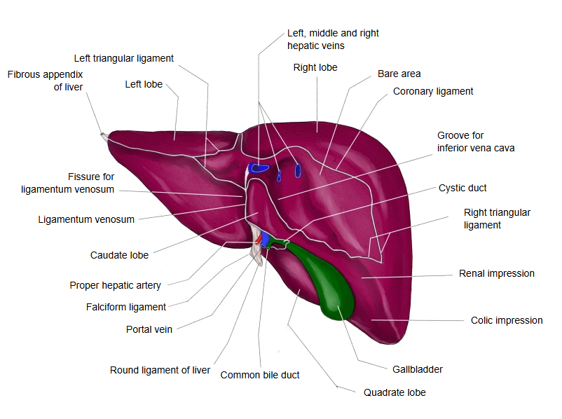
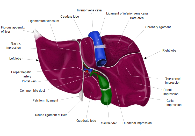
Over most of its surface the liver is covered by smooth peritoneum and slides freely against the diaphragm. In a narrow posterior region, however, the two are held together by fibrous connective tissue and hardly move. This region of the liver surface is the bare area (Latin area nuda, "naked area", because it has no peritoneal covering). It is roughly triangular, lies mostly on the right lobe, and is outlined by the upper and lower layers of the coronary ligament; the groove for the vena cava lies within it. In front, the upper layer of the coronary ligament continues as the falciform ligament; to the left and right the bare area narrows into the left and right triangular ligaments. The tip of the left triangular ligament forms a fibrous band joining the upper left end of the liver to the diaphragm, called the fibrous appendix of liver (appendix fibrosa hepatis).
· Relations: the inferior vena cava runs in its groove on the bare area, and the hepatic veins open into it here. The right suprarenal gland (whose impression lies partly on the bare area) and the upper pole of the right kidney lie close below.
· Lymph vessels from the bare area pass through the diaphragm to nodes in the thorax, and small veins here connect with veins of the diaphragm. These links allow infection or tumour to spread between the liver and the chest.
Reference websites
[Wikipedia]
· Bare area of the liver, Wikipedia.
· Coronary ligament, Wikipedia.
[Websites with illustrations]
· Bare area of liver (Kenhub)

· Liver, posterior view (Abdominal Key)
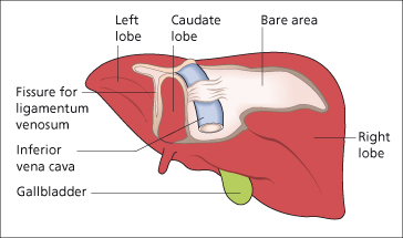
Where it is found
Related terms
- Groove for vena cava
- Suprarenal impression
- Visceral peritoneum
- Parietal peritoneum
- Right lobe of liver
- Liver
- Diaphragm
References and sources
- Further reading: Bare area of the liver, Wikipedia.
- Further reading: Coronary ligament of the liver, Wikipedia.
- Federative International Programme for Anatomical Terminology (FIPAT). Terminologia Anatomica, 2nd ed. (TA2), 2019.