Palpebral conjunctiva (Latin: tunica conjunctiva palpebrarum)
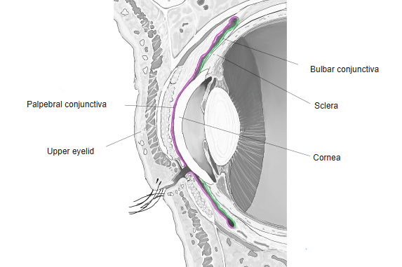
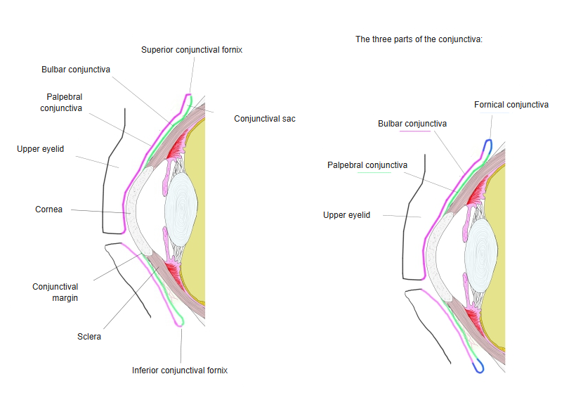
- Palpebral conjunctiva : covers the back of the eyelids; a membrane of two-layered or stratified columnar epithelium, rich in blood vessels.
- Bulbar conjunctiva : the part covering the eyeball; made of non-keratinised stratified squamous epithelium.
- Note : some sources, such as Wikipedia, add a third part, the fornical conjunctiva.
The palpebral conjunctiva has the following features.
· It is rich in blood vessels and firmly attached to the tarsal plates, where it shows small depressions (the conjunctival fossae).
· It is covered by two-layered or stratified columnar epithelium with goblet cells, on a loose, vascular lamina propria.
· It is highly vascular and its surface bears many small papillae. Older descriptions call it thick and opaque; over the tarsal plates, however, it is thin and firmly attached, and the tarsal glands show through it as yellowish streaks.
· Its deeper part contains lymphoid tissue. At the lid margin it meets the skin and is continuous with the lining of the ducts of the tarsal glands; through the lacrimal puncta and canaliculi it is continuous with the lining of the lacrimal sac and nasolacrimal duct. At the superior and inferior conjunctival fornices it folds back onto the eyeball and continues as the bulbar conjunctiva over the sclera.
· Its colour is a quick bedside check: it looks pale in anaemia.
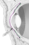 Front of eyeball (sagittal section)
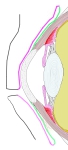 Conjunctiva (schematic)
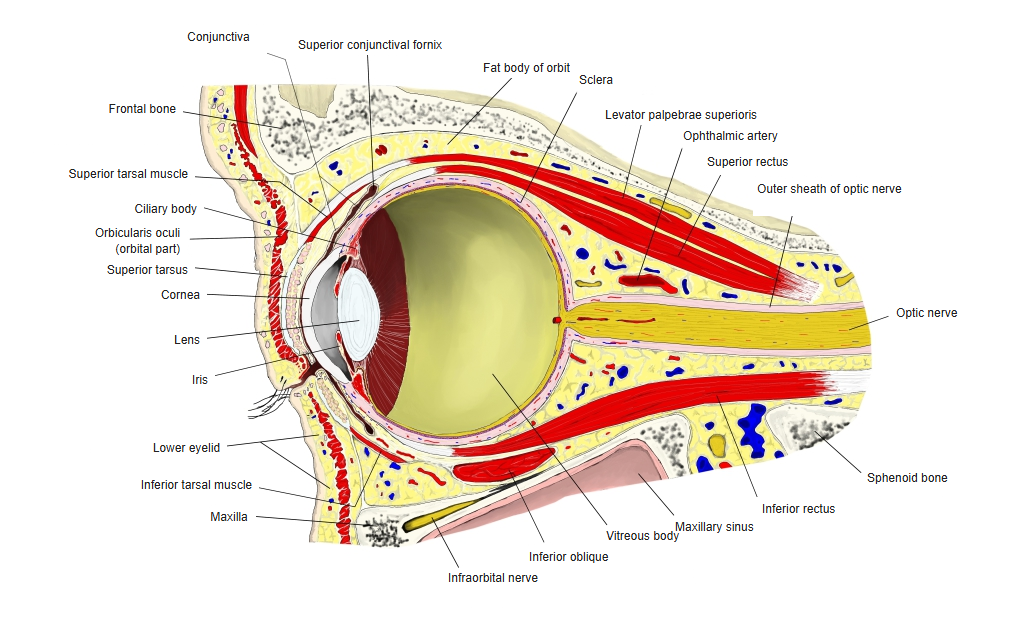
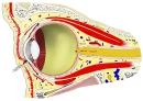 Orbit (sagittal section)
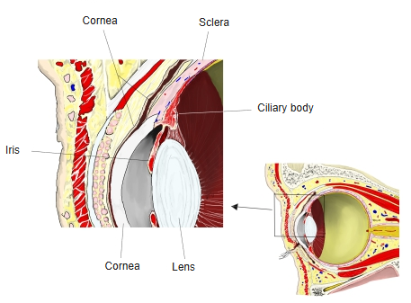
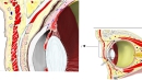 Front of orbit (sagittal section)
Reference websites
[Wikipedia]
· Conjunctiva, Wikipedia.
[Websites with illustrations]
· Parts of the conjunctiva (figure) (ctfassets.net)

· Eye and eyelid anatomy (illustration) (Science Photo Library)
· Cross-section of the eye and eyelids (figure) (VCA Hospitals)

Related terms
References and sources
- Further reading: Conjunctiva, Wikipedia.
- Federative International Programme for Anatomical Terminology (FIPAT). Terminologia Anatomica, 2nd ed. (TA2), 2019.