Peripheral process
Also called the peripheral axon of a sensory neuron
Overview
From the cell body of a pseudounipolar neuron a short process leaves and soon divides in two; the peripheral process is one of these two branches.
Cell body: lies in the dorsal root ganglion of a spinal nervePseudounipolar neuron: receives the signals from the skin and other sense organsPeripheral process: the long branch that runs to the skin and other sense organs
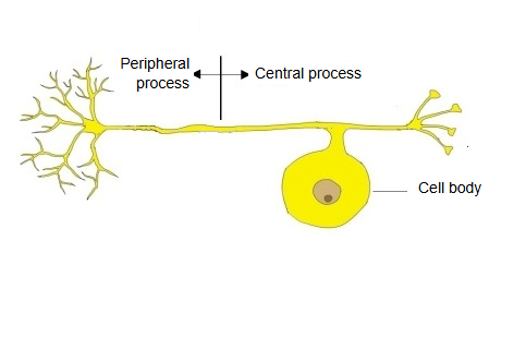
The peripheral process is sometimes called the "dendrite" of the pseudounipolar neuron, but in function it is an axon.
It conducts action potentials
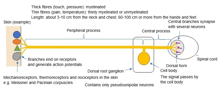
Peripheral processes are either myelinated or unmyelinated; when the peripheral process is myelinated (or unmyelinated), the central process is myelinated (or unmyelinated) too.
Thick fibres (touch, pressure) and the thin A-delta fibres (sharp pain, cold) are myelinated; the thinnest C fibres (dull pain, warmth) are unmyelinated
The length of the peripheral process differs greatly with the body region: from about 3 cm for the shortest to a metre or more for the longest.
Short: those receiving signals from the skin of the neck and chestLong: those from the skin of the fingers and toes
In the peripheral process the site where action potentials arise lies in the process itself; in this it differs greatly from other neurons.
In multipolar and bipolar neurons they arise at the axon hillock, between the cell body and the axon
· Sources differ on whether to call the peripheral process a dendrite or an axon; most current texts treat both processes as one axon divided into two branches.
Signal pathway
Signal pathway: receptor ⇒ ending of the peripheral process ⇒ impulse-generating zone ⇒ dorsal root ganglion ⇒ posterior horn of the spinal cord.
Receptor: mechanical, thermal and other stimuliEnding: receptor potentialImpulse-generating zone: action potentialsDorsal root ganglion to posterior horn: the central process
From the receptor to the spinal cord
Peripheral process
Receptor endingin skin, muscle and other tissues; mechanical, thermal or chemical stimuli open ion channels and produce a receptor potential
Trigger zonejust central to the ending; voltage-gated channels turn the receptor potential into action potentials
Cell body
Dorsal root ganglionaction potentials pass the cell body; no synapse here
Spinal cordenters through the dorsal root; synapses in the posterior horn
| Kind of sensation | Peripheral process | Fibre type | Examples of receptors |
|---|---|---|---|
| Touch (fine touch, vibration, pressure, position of the skin) | Myelinated | A-beta | Meissner corpuscles, Merkel discs, Pacinian corpuscles, Ruffini endings |
| Stretch and tension of muscles (deep sensation) | Myelinated | A-alpha (Ia, Ib) and A-beta | Muscle spindles, Golgi tendon organs |
| Sharp pain, cold | Myelinated (thin) | A-delta | Free nerve endings |
| Dull pain, warmth | Unmyelinated | C | Free nerve endings |
Main organs and tissues served by pseudounipolar neurons
| Organ or tissue | Stimuli sensed | Examples of receptors | |
|---|---|---|---|
| 1 | Skin | Touch, pain, temperature | Pacinian corpuscles, Meissner corpuscles, free nerve endings, TRP channels |
| 2 | Skeletal muscle | Stretch, contraction, pain | Muscle spindles (Ia fibres), nociceptors |
| 3 | Joints | Joint angle, pressure, pain | Receptors in the joint capsule, free nerve endings |
| 4 | Tendons | Muscle tension | Golgi tendon organs (Ib fibres) |
| 5 | Periosteum | Pain, pressure | Free nerve endings |
| 6 | Internal organs | Stretch, pain, chemical stimuli | Free nerve endings (visceral afferent fibres) |
| 7 | Blood vessels | Blood pressure, blood flow, chemical stimuli | Baroreceptors and chemoreceptors (carotid sinus, carotid body) |
Related terms
References and sources
- Further reading: Pseudounipolar neuron, Wikipedia.
- Further reading: Sensory neuron, Wikipedia.
- Federative International Programme for Anatomical Terminology (FIPAT). Terminologia Anatomica, 2nd ed. (TA2), 2019.