Pseudounipolar neuron
Also called the pseudounipolar nerve cell or the unipolar sensory neuron
- Overview
- Questions and answers
- Reference websites
- (Wikipedia, websites with illustrations)
- Related terms
Overview
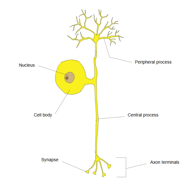
· In the embryo the cell starts with two processes, as a bipolar cell; later they come together near the cell body into one stalk, hence "pseudo"-unipolar.
· Of the two branches that divide just after leaving the cell body, one is called the peripheral process and the other the central process.
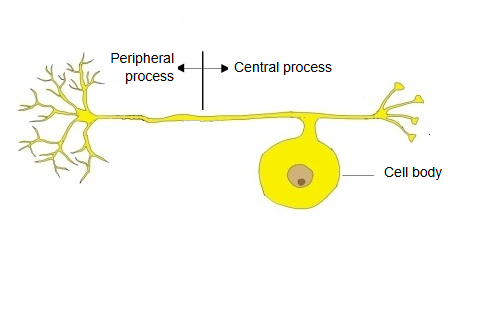
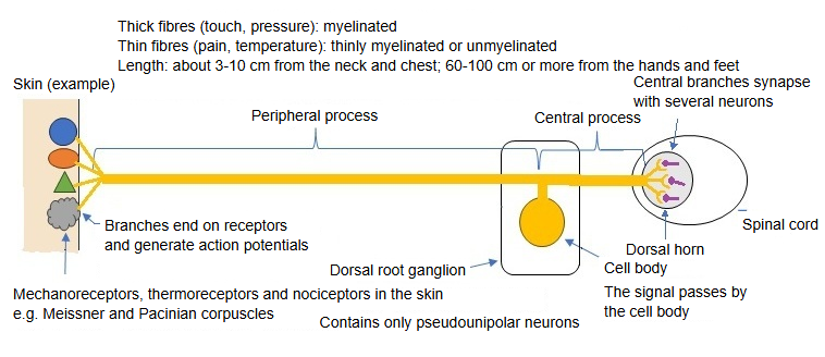
· Their cell bodies lie in the spinal ganglia (dorsal root ganglia) and in the sensory ganglia of most cranial nerves, such as the trigeminal ganglion and the sensory ganglia of the facial, glossopharyngeal and vagus nerves. The mesencephalic nucleus of the trigeminal nerve is the only group inside the central nervous system. The spiral and vestibular ganglia contain bipolar neurons instead.
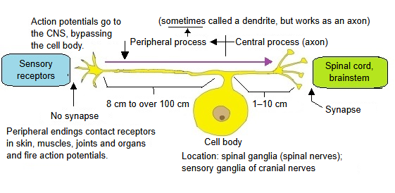
· The peripheral process runs to receptors in the skin, muscles, joints and organs; the central process enters the spinal cord or brainstem and synapses there. Both processes conduct action potentials and may be myelinated, so they are treated as two branches of one axon, forming primary afferent fibres; the signal can pass the cell body without going through it. Some texts call the peripheral process a dendrite because it carries signals towards the cell body; sources differ.
nerve cells
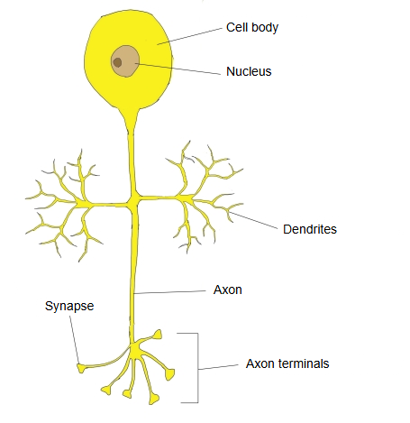
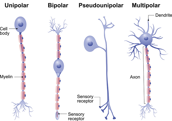
- Unipolar neuron : one process from the cell body. Found in the central nervous system of invertebrates; true unipolar neurons are not a usual type in humans.
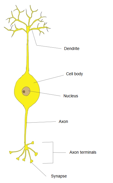
- Bipolar neuron : two processes from the cell body. Sites: the retina, the olfactory mucosa, and the ganglia of the vestibular and cochlear nerves. Only a small part of all neurons.
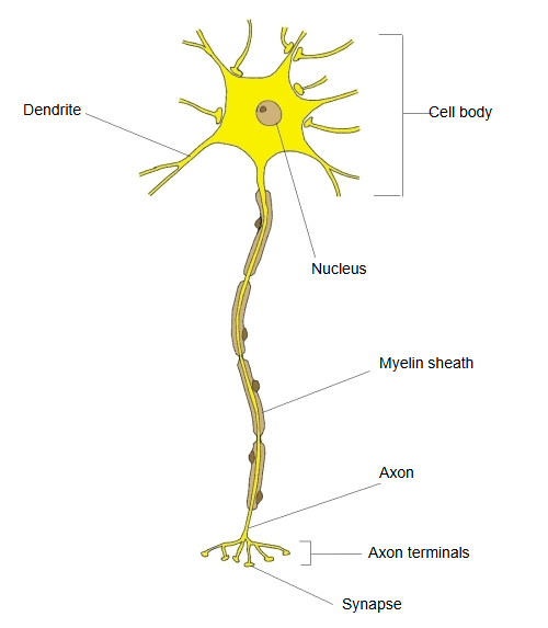
- Multipolar neuron : many processes from the cell body; almost all neurons of the central nervous system. Sites: the cortex of the brain, the spinal cord, the autonomic ganglia. By far the most common type.
- Pseudounipolar neuron : one process from the cell body that divides into two (peripheral and central process). The electrical signal does not have to pass through the cell body. Only a small part of all neurons.
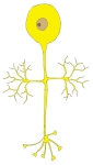 Unipolar neuron
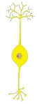 Bipolar neuron
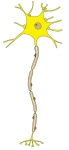 Multipolar neuron
 Pseudounipolar neuron
Questions and answers
Q1: Are there myelinated and unmyelinated pseudounipolar neurons?
A: Yes. The thick Aβ fibres (touch and pressure) are myelinated; the thin C fibres (pain, temperature, itch and others) are unmyelinated.
| Kind of sensation | Peripheral process | Fibre class | Examples |
|---|---|---|---|
| Touch (fine touch, vibration, pressure, position sense of the skin) | Myelinated | Aβ fibres | Meissner's corpuscles, Merkel discs, Pacinian corpuscles, Ruffini endings |
| Stretch and tension of muscles (deep sensation) | Myelinated | Aα (Ia, Ib) and Aβ fibres | Muscle spindles, Golgi tendon organs |
| Pain (sharp pain), cold | Myelinated | Aδ fibres | Free nerve endings |
| Pain (dull pain), warmth | Unmyelinated | C fibres | Free nerve endings |
Q2: Can myelinated and unmyelinated cells lie side by side (in the same ganglion)?
A: Yes. For example, a dorsal root ganglion of a lumbar nerve contains both myelinated cells with Aβ fibres (touch) and unmyelinated cells with C fibres (dull pain).
Q3: Is it easy to tell the peripheral process from the central process by their form?
A: No. The two have almost the same form and are hard to tell apart.
Q4: The peripheral process receives the information from the sense organs, so should it not be called a dendrite rather than an axon?
A: It is tempting to think "receives information = dendrite", but the right way to think is "axon = generates and conducts action potentials".
· In the peripheral process, an action potential arises directly from the stimulus at the sense organ ⇒ a feature of an axon.
An ordinary dendrite only turns the input it receives into graded changes of potential and does not fire action potentials.
· The peripheral process is often covered by a myelin sheath ⇒ a feature of an axon.
Dendrites have no myelin sheath.
· The peripheral process has no synapse; it senses physical and chemical stimuli directly and fires ⇒ not a feature of a dendrite.
An ordinary dendrite receives synaptic input from other neurons.
Q5: How do the peripheral and central processes differ in length?
A: There is a clear difference: usually the peripheral process is far longer and the central process short.
Peripheral process: from the sensory receptor (in the skin, muscles and other parts) to the dorsal root ganglion of the spinal cord ⇒ several centimetres to tens of centimetres, and in the longest limbs about a metre.
Central process: from the dorsal root ganglion to the posterior horn of the spinal cord ⇒ much shorter, millimetres to centimetres.
Reference websites
[Wikipedia]
· Pseudounipolar neuron, Wikipedia.
· Dorsal root ganglion, Wikipedia.
[Websites with illustrations]
· Pseudounipolar neuron (illustration) (Science Photo Library)

· Myelinated pseudounipolar sensory neuron (icon) (BioRender)

· Types of neurons (figure) (Queensland Brain Institute)
Related terms
- Peripheral process
- Spinal ganglion
- Dorsal root ganglion
- Sensory ganglion
- Axon
- Dendrite
- Primary afferent fibers
References and sources
- Further reading: Pseudounipolar neuron, Wikipedia.
- Further reading: Dorsal root ganglion, Wikipedia.
- Federative International Programme for Anatomical Terminology (FIPAT). Terminologia Anatomica, 2nd ed. (TA2), 2019.