Tympanic membrane (Latin: membrana tympanica)
Also called the eardrum
- Overview
- Parts
- Relation to the external acoustic meatus
- Layers
- Terms related to the tympanic membrane
- Vessels and nerves
- Reference websites
- (Wikipedia, websites with illustrations)
- Where it is found
- Related terms
Overview
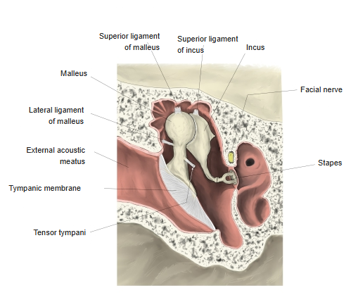
· The handle of the malleus is attached to its inner surface and pulls the centre inwards; the deepest point is the umbo. The tensor tympani, pulling on the malleus, tightens the membrane.
· Its vibrations pass along the auditory ossicles to the inner ear. A perforation or retraction of the membrane causes conductive hearing loss.
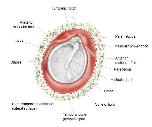
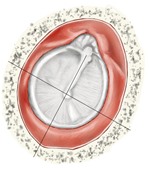
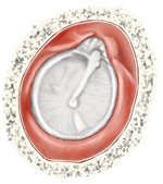 Right tympanic membrane (lateral surface)
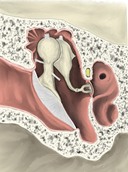 Right tympanic cavity (coronal section)
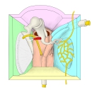 Right tympanic cavity (model)
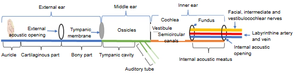
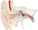 Right external ear to middle ear
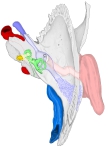 Right temporal bone (superior view)
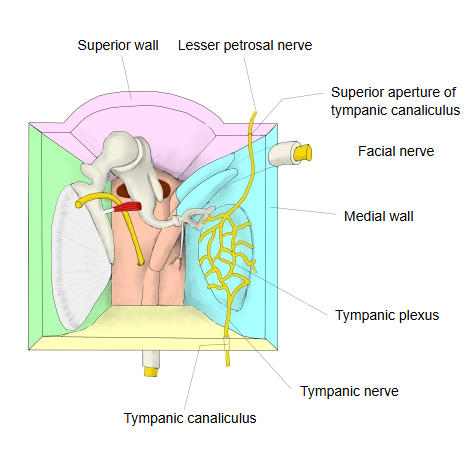
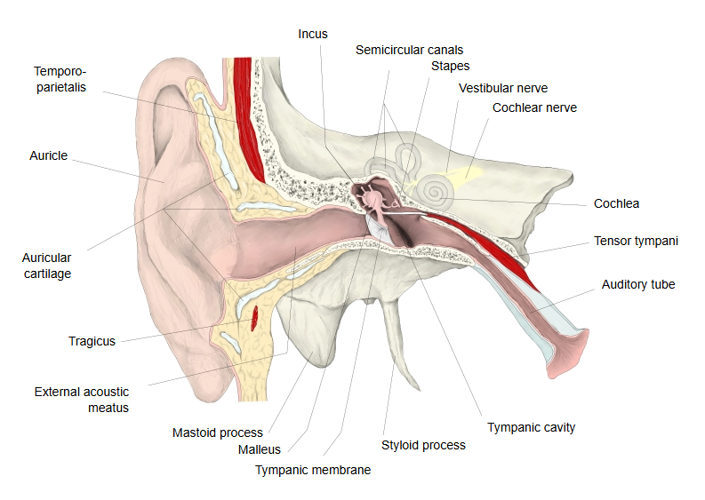
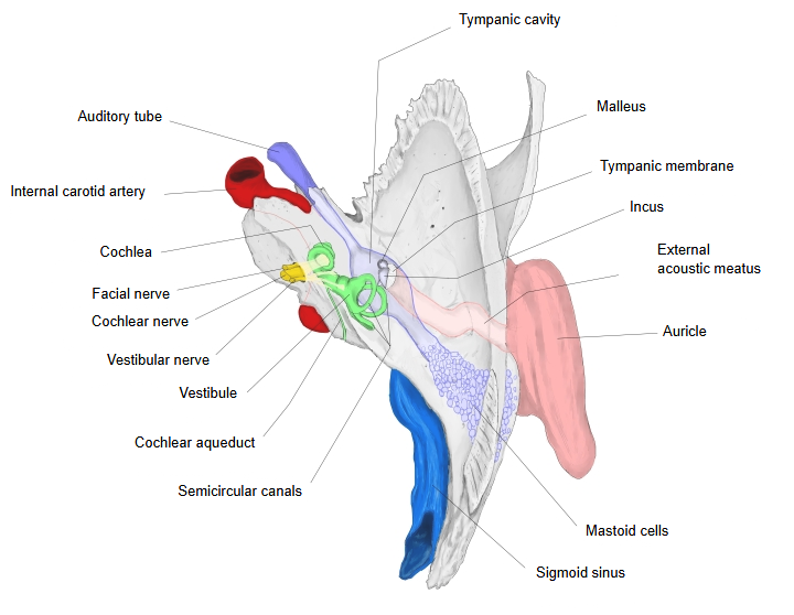
Parts
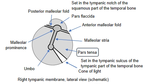
| Parts of the tympanic membrane | Pars flaccida | Flaccid part (Shrapnell's membrane). · The small, thin upper part of the membrane, above the malleolar prominence and the anterior and posterior malleolar folds. · It is set into the tympanic notch, the gap at the top of the tympanic ring of the temporal bone. |
| Pars tensa | Tense part. · The large remainder of the membrane; the attached handle of the malleus pulls it inwards into a funnel shape toward the tympanic cavity. · Its thickened rim, a fibrocartilaginous ring, is set into the tympanic sulcus of the tympanic part of the temporal bone. · A little below the centre, the tip of the handle of the malleus makes a depression, the umbo. |
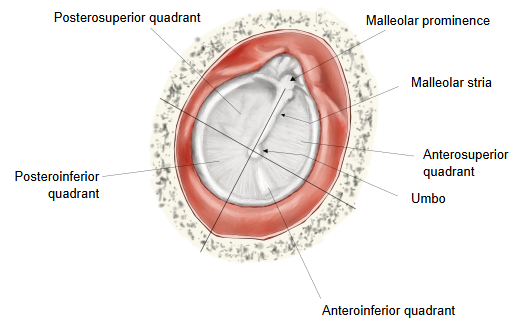
The quadrants are drawn as follows.
A line along the malleolar stria, continued downwards, and a line at right angles to it through the umbo divide the pars tensa into the anterosuperior, posterosuperior, anteroinferior and posteroinferior quadrants; the pars flaccida is counted as a fifth area. The incus and stapes lie behind the posterosuperior quadrant, so incisions into the membrane (myringotomy) are made in the inferior quadrants, from the anteroinferior to the posteroinferior quadrant.
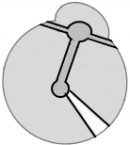 Right tympanic membrane (schematic)
 Clinical quadrants
Relation to the external acoustic meatus
In the adult it is set obliquely, facing laterally, forwards and downwards; its angle to the floor of the external acoustic meatus is given as roughly 40–55° (sources differ). Above, it passes into the roof of the meatus without a clear bend (an obtuse angle of about 140°); with the anterior wall it makes an acute angle of about 27°.
Layers
From the outside inwards, three layers are distinguished.
| 1 | Cutaneous layer (outer layer): continuous with the skin of the external acoustic meatus. | ||||
| a | Epidermis | A thin stratified squamous epithelium. Unlike ordinary skin, its surface (keratinized) cells are not shed on the spot but migrate outwards to the rim of the membrane. | |||
| b | Dermis | Connective tissue without papillae toward the epidermis; there are no sweat glands, sebaceous glands or hairs. | |||
| 2 | Lamina propria (middle, fibrous layer): tough fibrous connective tissue that forms the middle layer of the membrane. | ||||
| a | Outer radial fibre layer | Fibre bundles radiating from the umbo and the handle of the malleus; thickest in the centre. | |||
| b | Inner circular fibre layer | Fibre bundles running in rings around the umbo; thickest at the rim. In the pars flaccida, unlike the pars tensa, the fibres are sparse and run irregularly; this part reacts sensitively to changes of pressure in the tympanic cavity. | |||
| 3 | Mucous layer (inner layer): continuous with the mucosa of the tympanic cavity. | ||||
| a | Lamina propria of the mucosa | A thin, loose connective tissue layer on the outer side. | |||
| b | Simple squamous (to cuboidal) epithelium | On the inner side; it has no cilia, except where it passes into the mucosa of the tympanic cavity. | |||
Terms related to the tympanic membrane
- Anterior malleolar fold
- Cone of light
- Handle of malleus
- Lateral process of malleus
- Malleolar folds
- Malleolar prominence
- Malleolar stria
- Pars flaccida
- Pars tensa
- Posterior malleolar fold
- Tympanic notch
- Tympanic sulcus
- Umbo
Vessels and nerves
| 1 | Arteries | Inner surface: branches of the anterior tympanic artery. Outer surface: branches of the deep auricular artery. |
| Veins | Inner surface: the tympanic veins. Outer surface: veins in front of the auricle (anterior auricular veins). | |
| 2 | Lymph vessels | The subcutaneous lymphatic network drains to the lymph nodes behind the auricle; the mucosal network joins the lymph vessels of the tympanic cavity. |
| 3 | Nerves | Inner surface: branches of the tympanic plexus (from the glossopharyngeal nerve). Outer surface: shared with the external acoustic meatus: mainly the auriculotemporal nerve, with the auricular branch of the vagus nerve. |
Reference websites
[Wikipedia]
· Eardrum, Wikipedia.
[Websites with illustrations]
· Tympanic membrane: anatomy, structure and function (Kenhub)
· The middle ear (TeachMeAnatomy)
· Anatomy of the ear (Blausen Medical) (Wikimedia Commons)

Where it is found
Related terms
References and sources
- Further reading: Eardrum, Wikipedia.
- Federative International Programme for Anatomical Terminology (FIPAT). Terminologia Anatomica, 2nd ed. (TA2), 2019.