Perpendicular plate (Latin: lamina perpendicularis)
· The name "perpendicular plate" is used for two thin, vertical plates of bone: this one, part of the ethmoid bone in the nasal septum, and the perpendicular plate of the palatine bone in the side wall of the nasal cavity (see below).
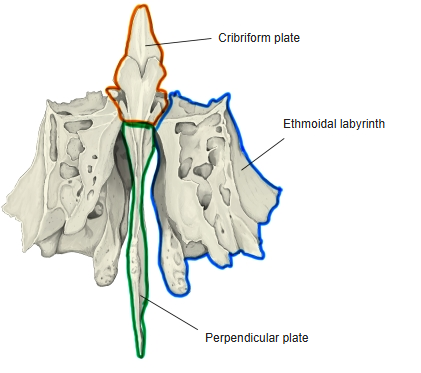
- Cribriform plate : the horizontal bony plate in the middle.
- Perpendicular plate : the plate that projects downwards from the underside of the cribriform plate.
- Ethmoidal labyrinth : the parts on both sides of the perpendicular plate that hang down from the cribriform plate.
the perpen-
dicular plate
- Superior border : joins the nasal bones and the frontal bone (nasal spine).
- Anterior border : joins the septal nasal cartilage.
- Inferior border : in front it joins the septal nasal cartilage, behind it joins the vomer.
- Posterior border : joins the sphenoid bone (sphenoidal crest of the body).
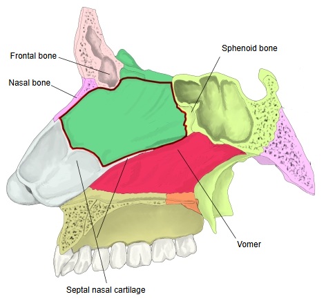
· Texts name the edges of the plate differently: many English texts call the edge that meets the frontal spine and the nasal bones the anterior border, the edge that meets the sphenoidal crest and the vomer the posterior border, and the thicker edge that holds the septal nasal cartilage the inferior border.
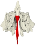 Ethmoid bone: anterior view
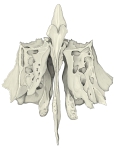 Ethmoid bone: anterior view, main features
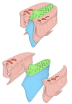 Ethmoid bone: its parts
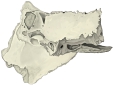 Ethmoid bone: lateral view
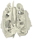 Ethmoid bone: second view
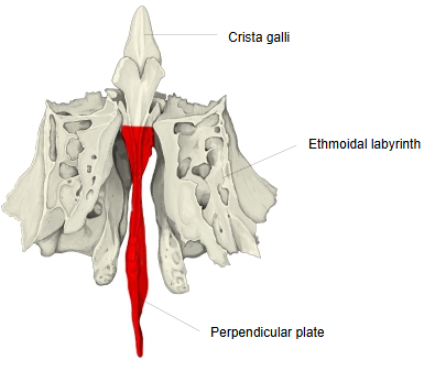
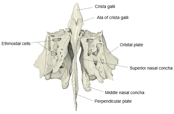
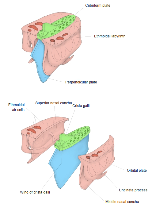
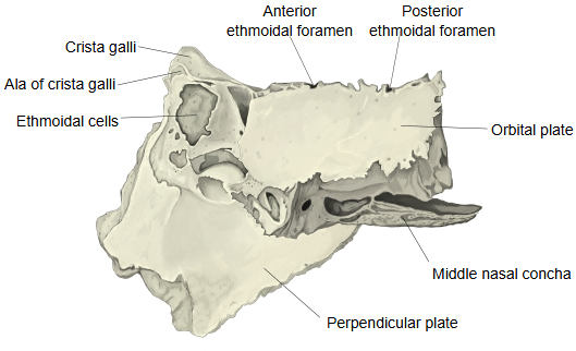
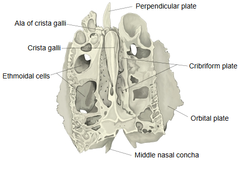
The perpendicular plate descends from the midline of the underside of the cribriform plate and stands vertically, almost in the median plane, as a thin bony plate of irregular triangular shape. It forms the upper front part of the bony nasal septum and is often bent a little to one side. The upper part of its front edge meets the nasal spine of the frontal bone and the crest of the nasal bones; its lower edge, the thickest, holds the septal nasal cartilage. Its back edge meets the sphenoidal crest on the body of the sphenoid above and the vomer below. Above the cribriform plate it continues upwards as the crista galli. The upper part of both surfaces carries many small grooves and canals, leading down from the foramina of the cribriform plate, for the filaments of the olfactory nerves.
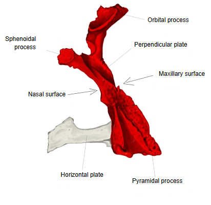
Perpendicular plate of palatine bone
- Perpendicular plate : the upright plate; it has two surfaces (nasal surface and maxillary surface) and three processes.
- Orbital process : projects upwards and laterally from the upper end of the perpendicular plate and forms a very small part of the orbit.
- Sphenoidal process : projects upwards and medially from the upper end of the perpendicular plate and joins the sphenoid bone.
- Pyramidal process : projects backwards and laterally from the junction of the perpendicular and horizontal plates (the lower back corner).
- Horizontal plate : the horizontal plate; it has two surfaces (nasal surface = superior surface, palatine surface = inferior surface).
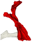 Right palatine bone (posterior view)
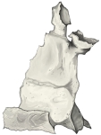 Right palatine bone (nasal surface)
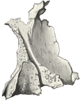 Right palatine bone (maxillary surface)
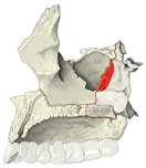 Maxilla and palatine bone (nasal surface)
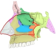 Nasal cavity (lateral wall)
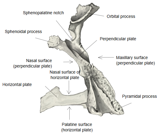
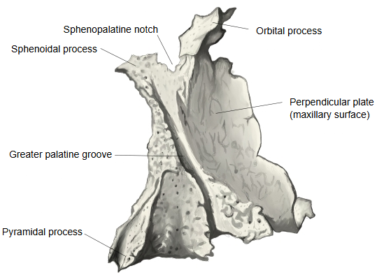
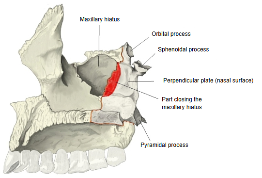
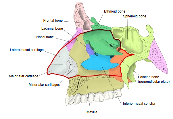
· At its upper end the orbital and sphenoidal processes are separated by the sphenopalatine notch.
· On its maxillary surface a groove, together with the maxilla, forms the greater palatine canal.
· Both perpendicular plates (of the ethmoid and of the palatine bone) are thin and fragile, so they are often broken in dried skulls.
Reference websites
[Wikipedia]
· Perpendicular plate of ethmoid bone, Wikipedia.
· Perpendicular plate of palatine bone, Wikipedia.
· Palatine bone, Wikipedia.
[Websites with illustrations]
· Ethmoid bone: anatomy, borders and development (Kenhub)
· Ethmoid bone from behind (Gray's Anatomy, fig. 151) (Wikimedia Commons)

· Palatine bone: plates, borders, processes, articulations (Kenhub)
· Left palatine bone, posterior aspect (Gray's Anatomy, fig. 169) (Wikimedia Commons)

Where it is found
Related terms
- Cribriform plate
- Crista galli
- Ethmoidal labyrinth
- Nasal septum
- Horizontal plate
- Pyramidal process
- Orbital process
References and sources
- Further reading: Perpendicular plate of ethmoid bone, Wikipedia.
- Further reading: Palatine bone, Wikipedia.
- Federative International Programme for Anatomical Terminology (FIPAT). Terminologia Anatomica, 2nd ed. (TA2), 2019.