Medullary rays (Latin: radii medullares)
Also called the medullary ray or the pars radiata or the pyramids of Ferrein
- Overview
- Comparison of contents
- Share of the contents
- Reference websites
- (Wikipedia, websites with illustrations)
- Related terms
Overview
· A medullary ray is a bundle of renal tubules and other straight tubes that runs radially in the renal cortex from the renal medulla towards the surface of the cortex. It is also called Ferrein's pyramid (pars radiata).
· It is medullary tissue that enters the cortex like a band, and it does not reach the renal capsule.
· It contains the straight parts of the proximal tubule and the distal tubule (the straight tubules; the distal one is the thick ascending limb) and the cortical collecting ducts. It contains no renal corpuscles.
· Between neighbouring medullary rays lies the cortical labyrinth, where the renal corpuscles and the convoluted renal tubules gather; the renal cortex consists of the medullary rays and the cortical labyrinth.
· One medullary ray with the nephrons that drain into it forms a cortical lobule (renal lobule); the interlobular arteries and veins run between neighbouring lobules.
· In microscope sections a medullary ray is seen as a row of straight tubules without renal corpuscles, which distinguishes it from the cortical labyrinth.
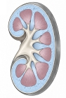 Renal cortex and renal medulla
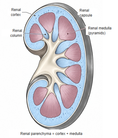
cortex
- Cortical labyrinth : renal corpuscles, proximal convoluted tubules, distal convoluted tubules, connecting tubules, juxtaglomerular apparatus, interlobular arteries and veins, afferent and efferent arterioles, capillariesabout 70–75%
- Medullary rays : proximal straight tubules, thick ascending limbs of the loop of Henle, collecting ducts, capillariesabout 25–30%
The percentages are approximate and differ between sources.
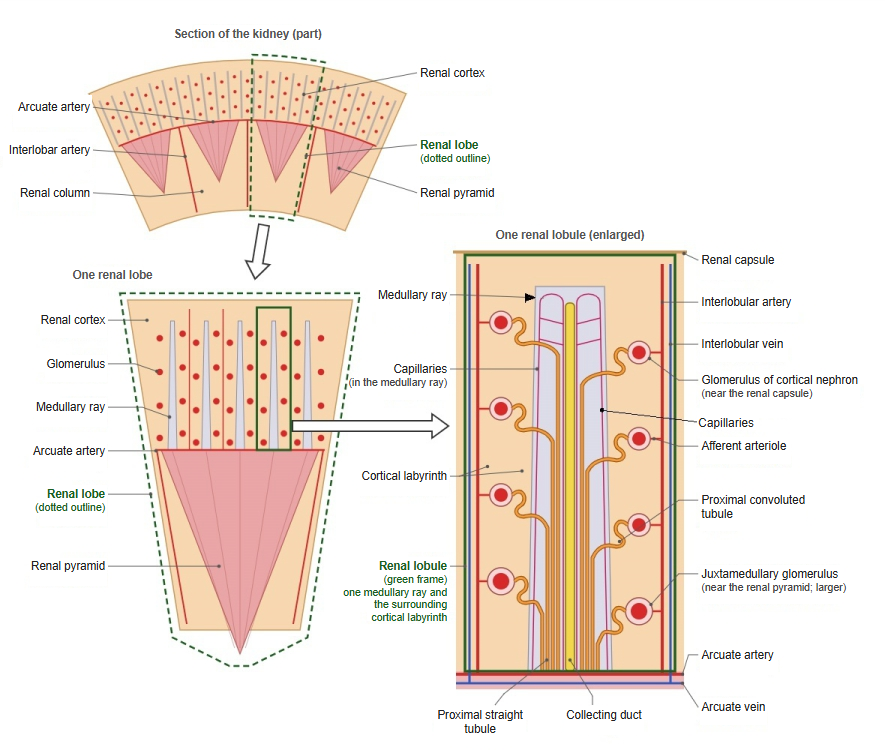
⇒ A more detailed diagram of the renal cortex and the medullary rays:
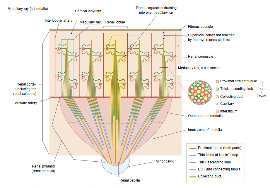
Medullary ray = a bundle of straight tubes (collecting ducts, proximal straight tubules and thick ascending limbs) that runs from the base of the renal pyramid into the cortex.
· It does not reach the fibrous capsule: the superficial layer of the cortex is cortical labyrinth only (cortex corticis).
· Each ray receives the tubules of many renal corpuscles (nephrons) of the cortical labyrinth around it; the unit centred on one ray is the renal lobule.
· Many rays arise from the base of each pyramid (the figure shows five).
· The tubes of a ray continue into the medulla of the pyramid; the loops of Henle turn in the medulla, and the collecting ducts converge on the renal papilla.
Comparison of contents
The table below compares the contents of the cortical labyrinth and the medullary rays.
| Component | Cortical labyrinth | Medullary ray |
|---|---|---|
| Location and appearance | Between the medullary rays. Renal corpuscles lie in it and the tubules are coiled. | Rays running from the medulla toward the capsule. Straight tubules run in bundles. |
| Nephron and collecting duct | ||
| Renal corpuscle (glomerulus + Bowman's capsule) | ○ All renal corpuscles lie here | – None |
| Proximal tubule | ○ Convoluted part | ○ Straight part (runs down into the medulla) |
| Distal tubule (straight part = thick ascending limb) | – None | ○ Rises from the medulla (thick ascending limb) |
| Distal tubule (convoluted part) | ○ Convoluted part; where it touches its own renal corpuscle it forms the macula densa | – None |
| Connecting tubule | ○ Joins the distal tubule to the collecting duct | – None |
| Collecting duct | Only its first part, where the connecting tubules join; it then turns into the medullary ray | ○ Cortical collecting duct (runs down into the medulla) |
| Blood vessels and other structures | ||
| Interlobular arteries and veins | ○ Run straight toward the capsule (between the lobules) | – None |
| Afferent and efferent arterioles | ○ Enter and leave the vascular pole of the renal corpuscle | – None |
| Juxtaglomerular apparatus | ○ Macula densa, juxtaglomerular cells, extraglomerular mesangial cells | – None |
| Capillaries | ○ Network of peritubular capillaries around the convoluted tubules | ○ Long, narrow capillary meshes along the straight tubules |
Note: one medullary ray with the cortical labyrinth around it forms one renal lobule.
Note: the medullary rays are bundles of straight tubules (straight parts and collecting ducts) that continue from the medulla into the cortex.
Share of the contents
By area, the largest part of a medullary ray is taken up by the straight parts of the proximal tubules.
A simple overview, in order of the space they take up:
| Structure | Size | Notes | |
|---|---|---|---|
| 1 | Proximal straight tubule | The thickest tube | One per nephron |
| 2 | Thick ascending limb | Thinner | About as many as the proximal straight tubules, but thinner |
| 3 | Collecting duct | Between the two | Fewer: each one receives the urine of several nephrons |
| 4 | Capillaries and interstitium | Very thin | Only fill the spaces between the tubes |
Most of the space is taken up by the tubes; between them there are only capillaries and a little interstitium. By number, proximal straight tubules and thick ascending limbs are about equal and far outnumber the collecting ducts; by area, the thick proximal straight tubules exceed the thick ascending limbs. The proportions in humans have not been measured.
Reference websites
[Wikipedia]
· Medullary ray (anatomy), Wikipedia.
· Renal cortex, Wikipedia.
[Websites with illustrations]
· Tubules and collecting system (Digital Histology)
· Urinary system overview (Digital Histology)
· Plate 12.232 Kidney: Cortex (Atlas of Microscopic Anatomy)
· Simplified structure of a kidney lobe (Wikimedia Commons)

Related terms
- Cortical labyrinth
- Renal sinus
- Renal parenchyma
- Kidney
- Interlobular arteries of kidney (cortical radiate arteries)
- Fibrous capsule of kidney
- Urinary system
References and sources
- Further reading: Medullary ray (anatomy), Wikipedia.
- Further reading: Renal cortex, Wikipedia.
- Federative International Programme for Anatomical Terminology (FIPAT). Terminologia Anatomica, 2nd ed. (TA2), 2019.