Elastic cartilage (Latin: cartilago elastica)
Also called the yellow cartilage
- Overview
- Composition
- Arrangement of the cells
- Where it is found
- Comparison of the three types of cartilage
- Related terms
Overview
Two views are found on which type of cartilage is the second most plentiful after hyaline cartilage:
Elastic cartilage: it is limited to the auricle, the external acoustic meatus, the auditory tube, the epiglottis and small laryngeal cartilages such as the cuneiform and corniculate cartilages, but it is spread more widely than fibrocartilage, so its total amount is thought to be larger.
Fibrocartilage: it is placed where the body bears heavy loads and strong twisting forces, such as the intervertebral discs, so its total amount is thought to be much larger than that of elastic cartilage.
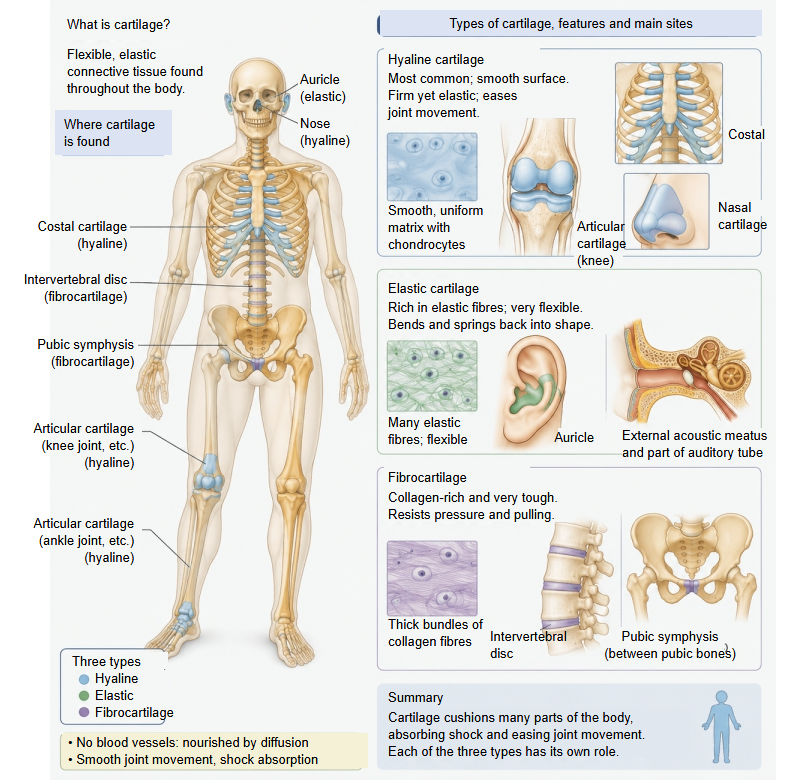
Like other cartilage it has no blood vessels or nerves; its cells are nourished by diffusion from the vessels of the perichondrium, which covers it everywhere. Unlike hyaline cartilage, it hardly ever calcifies or ossifies with age.
Composition
The word "rich" is used above, but the share of elastin in the whole tissue is not known. In the other two types of cartilage, however, there is none or only a negligible amount.
| Component | Share (approx.) | Role and features | ||
|---|---|---|---|---|
| 1 | Chondrocytes | 2-5% (5-10% in some sources) | Packed more densely than in the other two types of cartilage. Often several crowd into one lacuna. They actively secrete the extracellular matrix below. | |
| 2 | Extracellular matrix | Water | 60-70% | Tissue fluid that seeps slowly in from the vessels of the perichondrium. It carries nutrients and oxygen and gives the tissue its flexibility. |
| 3 | Elastic fibres (mainly elastin) | the rest (about 30-40%); estimates for the elastic fibres alone vary widely (about 15-35%) | The most typical feature of elastic cartilage (a simple protein). They form a dense mesh that gives great stretch and recoil. | |
| 4 | Collagen (= collagen fibres) | Mainly type II collagen. Together with the mesh of elastic fibres it forms the framework of the whole tissue. | ||
| 5 | Proteoglycans | Sugar chains such as chondroitin sulfate bound to a core protein. Their strong negative charge draws in and holds water. | ||
| 6 | Others | Link proteins, fibronectin and others, which bind the fibres, proteoglycans and cells to each other. | ||
Arrangement of the cells
An illustration of the microscopic structure (made with the help of AI):
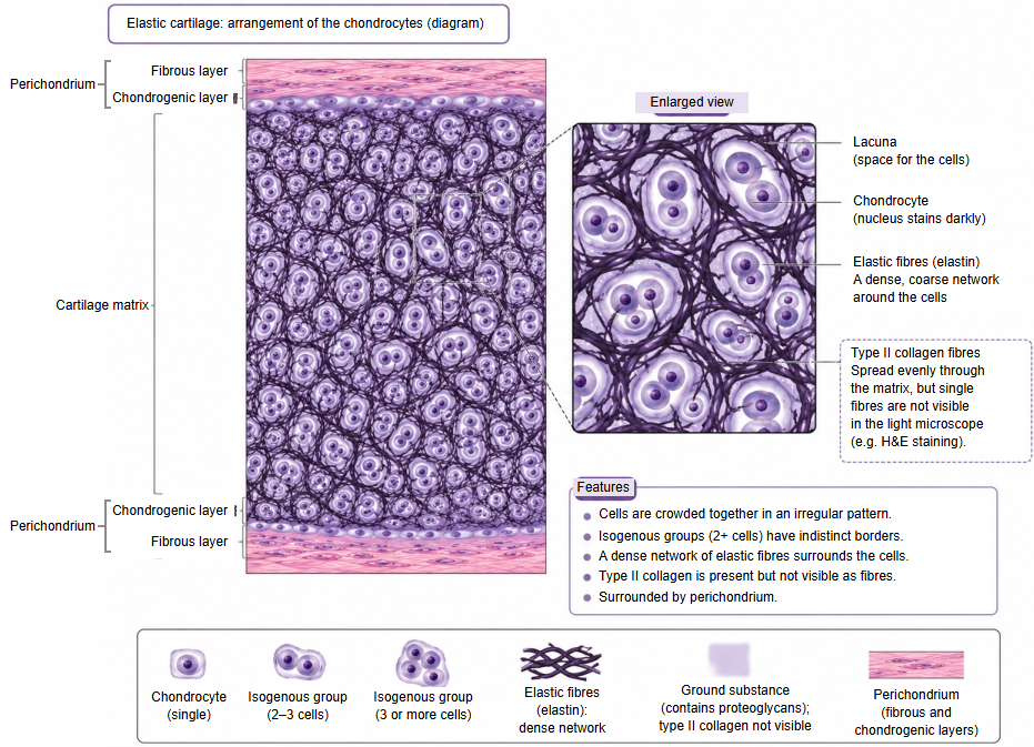
The cells make up only a few per cent of elastic cartilage, so why do they seem to fill the picture? The answer lies in the difference between weight and volume.
Volume: chondrocytes are large cells swollen with water, while the elastin and collagen around them are thin threads woven into a mesh. The swollen cells take up most of the field and the fibres run through the gaps between them, so under the microscope the tissue looks full of cells.
Weight: figures of a few per cent describe the cells' share of the tissue's mass; the solid parts of the cells weigh little compared with the collagen, elastin and other components of the matrix.
In hyaline cartilage, too, the cells are only a few per cent of the tissue, but the matrix between them is wide, so the cells look like scattered islands. In elastic cartilage the spaces between the cells are narrow, and the cells look densely crowded.
So the low percentage and the picture of crowded cells are both correct: the illustration shows how the tissue looks by volume under the microscope.
Where it is found
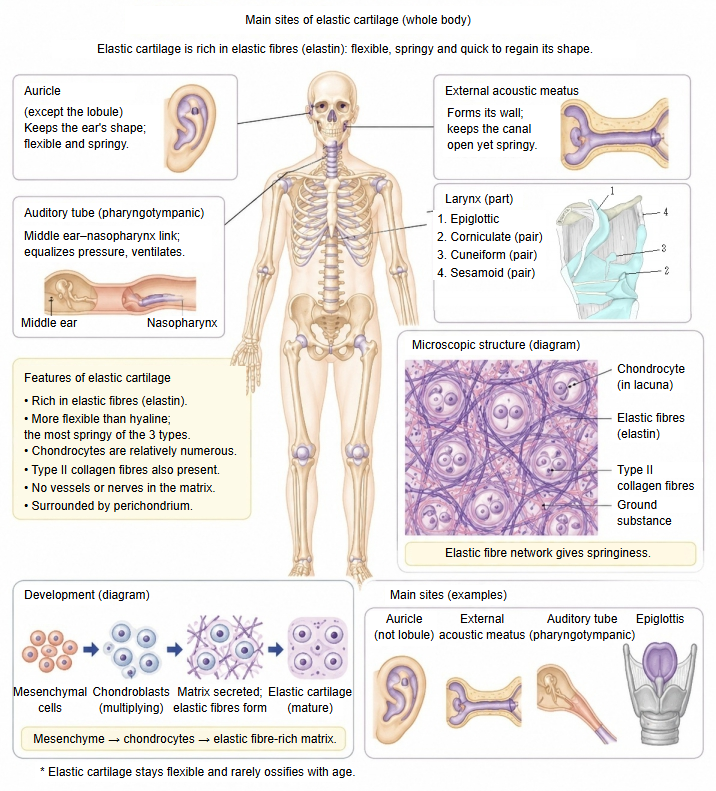
Elastic cartilage is found where the body needs a part that keeps its shape but bends and at once springs back.
| Site | Description | |
|---|---|---|
| 1 | Auricle | The cartilage of the outer ear (auricular cartilage); the earlobe itself has no cartilage. It lets the ear fold right over, for example when lying on one's side, and spring back at once without damage. |
| 2 | External acoustic meatus | The cartilage of the outer third of the ear canal (its cartilaginous part). It gives way when an ear pick or earphone is pushed in, so it does not hurt, yet keeps the canal from collapsing. |
| 3 | Part of the laryngeal cartilages | Of the laryngeal cartilages, the thyroid and cricoid cartilages and most of the arytenoid cartilage are hyaline; the unpaired epiglottic cartilage, the paired corniculate and cuneiform cartilages, the sesamoid cartilages and the vocal process and apex of each arytenoid are elastic cartilage. |
| 4 | Auditory tube | The cartilage of the tube that joins the middle ear to the throat. Usually closed, it opens easily when we swallow, and so equalizes the air pressure in the ear. |
Comparison of the three types of cartilage
A short comparison of the three types of cartilage:
| Feature | Hyaline cartilage | Elastic cartilage | Fibrocartilage | |
|---|---|---|---|---|
| 1 | Appearance | Translucent and smooth; fairly firm and resilient. | Yellowish and very elastic; springs back strongly. | Whitish and very tough; resists compression and pulling. |
| 2 | Cells | about 3-5% | about 2-5% (10% in some sources) | about 1-2% |
| 3 | Water | the most (about 70-80%) | about 60-70% | the least (about 50-65%) |
| 4 | Fibres | Type II collagen | Elastic fibres (elastin fibres) + type II collagen | Thick bundles of type I collagen + some type II |
| 5 | Perichondrium | Present (except on articular cartilage) | Present | Absent |
| 6 | Function | Support and shock absorption; smooth joint movement | Keeps flexibility and shape | Resists strong compression and pulling |
| 7 | Sites | Articular cartilage, costal cartilage, nasal cartilages, tracheal and bronchial cartilages, epiphyseal cartilage, part of the laryngeal cartilages (cricoid, thyroid, arytenoid) | Auricle, external acoustic meatus, auditory tube, part of the laryngeal cartilages (epiglottic, corniculate, cuneiform, sesamoid) | Intervertebral discs, pubic symphysis, menisci, articular discs, acetabular labrum and other labra, attachments of some tendons and ligaments to bone |
Related terms
- Elastic fibre
- Cartilage
- Hyaline cartilage
- Perichondrium
- Auricular cartilage
- Epiglottic cartilage
- Corniculate cartilage
- Cuneiform cartilage
References and sources
- Further reading: Elastic cartilage, Wikipedia.
- Further reading: Cartilage, Wikipedia.
- Federative International Programme for Anatomical Terminology (FIPAT). Terminologia Anatomica, 2nd ed. (TA2), 2019.