Fibrocartilage (Latin: cartilago fibrosa)
Also called the fibrous cartilage or the white fibrocartilage
- Overview
- Composition
- Arrangement of the cells
- Where it is found
- Comparison of the three types of cartilage
- Related terms
Overview
Two views are found on which type of cartilage is the second most plentiful after hyaline cartilage:
Elastic cartilage: elastic cartilage is found only in a few organs - the auricle, the external acoustic meatus, the auditory tube, the epiglottis and small laryngeal cartilages such as the cuneiform and corniculate cartilages - but in this view these sites together are spread more widely than those of fibrocartilage, so its total amount is thought to be larger.
Fibrocartilage: it is placed where the body bears weight and strong twisting forces, at the load-bearing pillars of the body, so its total amount is thought to be much larger than that of elastic cartilage.
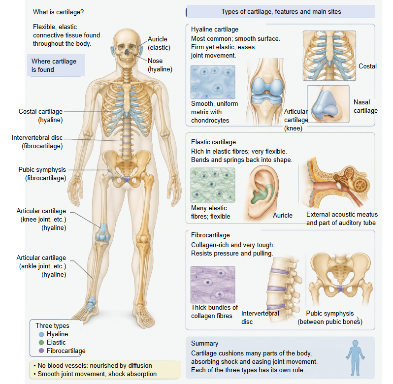
Like other cartilage it has no blood vessels, nerves or lymphatic vessels; unlike hyaline and elastic cartilage it usually has no perichondrium. It is a mixture of dense connective tissue and hyaline cartilage and often grades into one of them at its edges.
Composition
Type I and type II collagen fibres are not mixed at random: each has its own region.
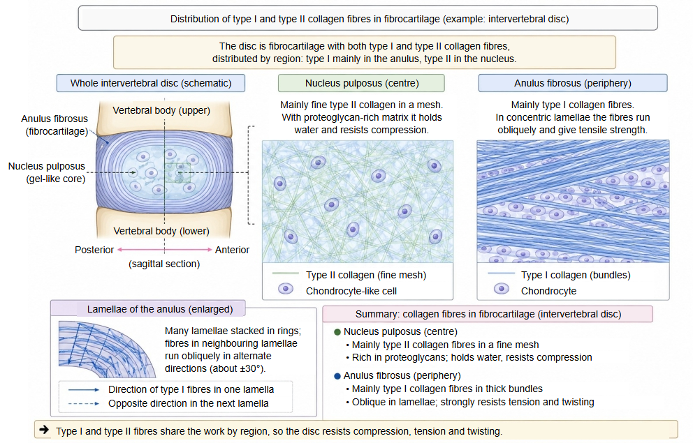
| Component | Share (approx.) | Role and features | ||
|---|---|---|---|---|
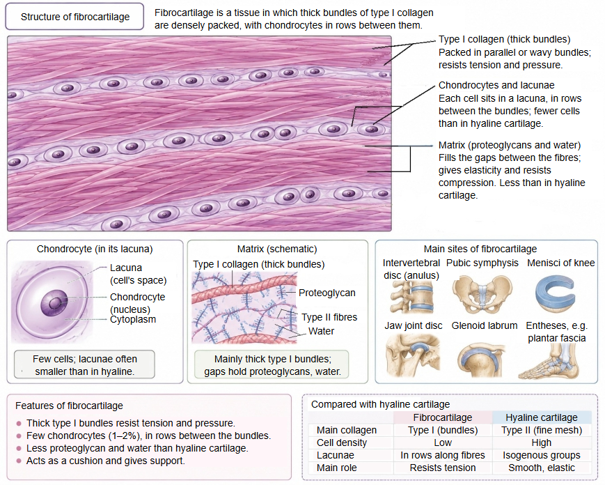
| 1 | Chondrocytes | 1-2% | The only cells: one or a few in each lacuna, often in a row like a string of beads. They maintain and repair the matrix and make collagen, proteoglycans and other components. | |
| 2 | Extracellular matrix | Water | 50-65% | Most of the matrix. It absorbs shocks under compression and gives elasticity; it is held in the tissue by the strong water-binding of the proteoglycans. |
| Collagen (= collagen fibres) | 25-35% | Mainly type I, which forms thick fibre bundles; the ratio to type II differs by site. In the intervertebral disc, type I is about 80-90% in the outer layer and about 40% in the inner layer. | ||
| Elastic fibres (elastin) | under 1% | Only traces of elastin molecules and microfibrils lie in the gaps of the tissue; the amount is negligible. | ||
| Proteoglycans | 2-5% | Giant molecules (such as aggrecan) fill the spaces between the collagen fibres. They hold much water and so help to resist compression. | ||
| Others | 1-3% | Adhesive glycoproteins (such as fibronectin and chondronectin) and other cell-attachment factors, growth factors, enzymes and others. | ||
Arrangement of the cells
Where it is found
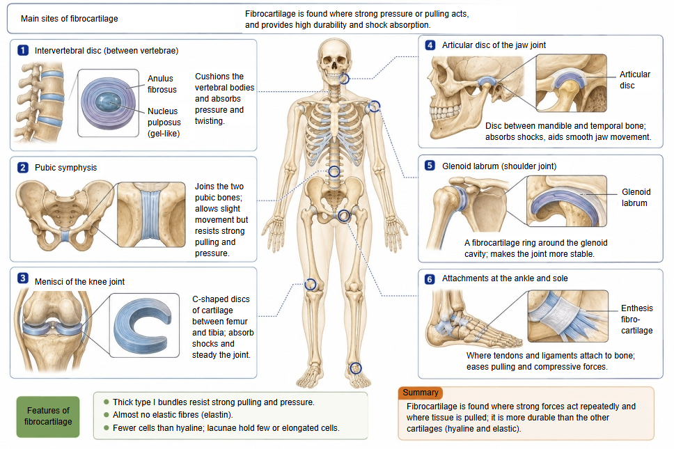
| Site | Examples | Main role and features | |
|---|---|---|---|
| 1 | Intervertebral disc (symphysis) | Anulus fibrosus | Mainly type I collagen; withstands compression, twisting and traction between the vertebrae. The nucleus pulposus is not fibrocartilage. |
| 2 | Pubic symphysis (symphysis) | Joint between the left and right pubic bones | Joins the pelvis and spreads the forces of walking and childbirth. |
| 3 | Manubriosternal joint (symphysis) | Joins the manubrium and the body of the sternum | Joins the front of the chest wall firmly, allows slight movement of the thorax in breathing and softens blows to the front of the chest. |
| 4 | Menisci | Medial and lateral menisci (knee joint) | Spread the load, absorb shocks and make the joint more stable. |
| 5 | Articular discs | Temporomandibular joint, sternoclavicular joint, distal radio-ulnar joint | Spread the load on the joint surfaces and improve their fit. |
| 6 | Articular labra | Glenoid labrum (shoulder), acetabular labrum (hip) | Deepen the socket and make the joint more stable. |
| 7 | Attachments of tendons and ligaments | Attachments of the calcaneal (Achilles) tendon, the patellar ligament, the rotator cuff and others | Pass the force from tendon or ligament to bone efficiently and spread the stress. |
Comparison of the three types of cartilage
A short comparison of the three types of cartilage:
| Feature | Hyaline cartilage | Elastic cartilage | Fibrocartilage | |
|---|---|---|---|---|
| 1 | Appearance | Translucent and smooth; fairly firm and resilient. | Yellowish and very elastic; springs back strongly. | Whitish and very tough; resists compression and pulling. |
| 2 | Cells | about 3-5% | about 2-5% (10% in some sources) | about 1-2% |
| 3 | Water | the most (about 70-80%) | about 60-70% | the least (about 50-65%) |
| 4 | Fibres | Type II collagen | Elastic fibres (elastin fibres) + type II collagen | Thick bundles of type I collagen + some type II |
| 5 | Perichondrium | Present (except on articular cartilage) | Present | Absent |
| 6 | Function | Support and shock absorption; smooth joint movement | Keeps flexibility and shape | Resists strong compression and pulling |
| 7 | Sites | Articular cartilage, costal cartilage, nasal cartilages, tracheal and bronchial cartilages, epiphyseal cartilage, part of the laryngeal cartilages (cricoid, thyroid, arytenoid) | Auricle, external acoustic meatus, auditory tube, part of the laryngeal cartilages (epiglottic, corniculate, cuneiform, sesamoid) | Intervertebral discs, pubic symphysis, menisci, articular discs, acetabular labrum and other labra, attachments of some tendons and ligaments to bone |
Related terms
- Cartilage
- Hyaline cartilage
- Elastic cartilage
- Chondrocyte
- Intervertebral disc
- Pubic symphysis
- Articular disc
- Acetabular labrum
References and sources
- Further reading: Fibrocartilage, Wikipedia.
- Further reading: Cartilage, Wikipedia.
- Federative International Programme for Anatomical Terminology (FIPAT). Terminologia Anatomica, 2nd ed. (TA2), 2019.