Antibody
Also called the immunoglobulin (Ig)
- Overview
- Structure
- How antibodies are made
- Classes
- Where antibodies are found
- Functions
- Reference websites
- (Wikipedia)
- Related terms
Overview
· B lymphocytes are also called B cells and are one kind of lymphocyte.
· B lymphocytes wait in the secondary lymphoid organs, such as the spleen and the lymph nodes. Only when a foreign substance has entered the body and they have received antigen presentation do they change into plasma cells and produce antibodies.
cytes
Humoral immunity
Structure
Chemically, an antibody is a glycoprotein: a protein built of amino acids, with sugar chains attached. The molecule is Y-shaped.
1. An antibody consists of two identical heavy chains (H chains) and two identical light chains (L chains), which together form the Y shape. Each light chain is joined to a heavy chain, and the two heavy chains to each other, by disulfide bonds (S-S bonds).
2. The tips of the two arms of the Y bind the antigen; their shape differs from antibody to antibody according to the antigen, and they are called the variable regions. The rest of the molecule has the same structure in all antibodies of a class and is called the constant region. Each arm (the variable region with the neighbouring constant parts of one heavy and one light chain) is called the Fab region (antigen-binding fragment); the stem, made of the constant parts of the two heavy chains, is the Fc region, which binds immune cells and complement.
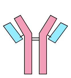 Heavy and light chains
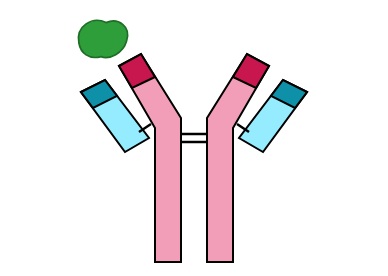
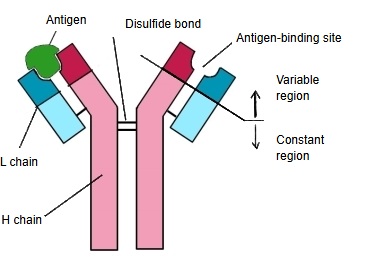 Parts of an antibody
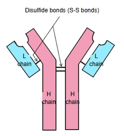
How antibodies are made
Antibodies are produced by the B cells (B lymphocytes), one kind of lymphocyte. Each B cell and its descendants (a clone) make antibodies against one antigen only. For example, when an adenovirus, a common cause of colds, enters the body, the B cells whose antibodies fit that virus are activated and multiply; their antibodies do not act against other viruses or toxins.
Because the bacteria, viruses and toxins that can enter the body are of an enormous number of kinds, the body keeps an equally enormous variety of B cell clones, each with its own antibody.
The figure below shows, in simple form, how B lymphocytes arise in the primary lymphoid organ (bone marrow) and are activated in the secondary lymphoid organs.
Life of a B cell
The bone marrow cells come mainly from the pelvis, vertebrae, sternum and ribs; the immature B cells are carried in the blood and lymph to the secondary lymphoid organs (spleen, lymph nodes and others).
Classes
Human antibodies (immunoglobulins, Ig) are divided into the following five classes.
| 1 | IgG | Immunoglobulin G. The most abundant class in the blood plasma, found both inside and outside the blood vessels; provides most antibody-based immunity and is the only class that crosses the placenta to the fetus. |
| 2 | IgM | Immunoglobulin M. The first class made in an infection, so it carries the early antibody response; the secreted form is a pentamer of five units and stays mainly in the blood. A single-unit form sits on the surface of B cells. |
| 3 | IgA | Immunoglobulin A. Protects the mucous membranes of the gut, airways and urogenital tract; the secreted form is a dimer of two units. |
| 4 | IgD | Immunoglobulin D. Present in very small amounts; works mainly as an antigen receptor on the surface of B cells that have not yet met an antigen. |
| 5 | IgE | Immunoglobulin E. The least abundant class; takes part in the defence against parasites and in allergy, by making mast cells and basophils release histamine. |
Where antibodies are found
1. Blood plasma and serum: the main place, holding mostly IgG, with IgM, IgA and others.
2. Mucosal secretions (saliva, tears, intestinal juice, breast milk and others): mainly IgA, which acts as local immunity; the amount is smaller than in the blood.
3. Lymph and interstitial fluid: antibodies that have passed out of the plasma.
4. On the cell membrane of B cells: some antibodies (IgM and IgD) are bound to the membrane as antigen receptors; their amount is very small.
Functions
Antibodies act mainly in the following three ways.
| 1 | Opsonization | Antibodies bind the antigen and coat it, which strengthens its ingestion (phagocytosis) by macrophages and neutrophils. |
| 2 | Complement activation | When antibodies bind bacteria that have entered the body, they activate complement (a group of blood proteins), which damages the cell membrane of the bacteria and makes them burst. |
| 3 | Neutralization | Viruses and toxins attach to cells by part of their structure before they enter the cells or harm them. Antibodies that bind that part first block the attachment and so reduce the harm. |
Antibodies in brief
Reference websites
[Wikipedia]
· Antibody, Wikipedia.
Related terms
References and sources
- Further reading: Antibody, Wikipedia.
- Federative International Programme for Anatomical Terminology (FIPAT). Terminologia Anatomica, 2nd ed. (TA2), 2019.