First rib (Latin: costa prima; costa I)
Position
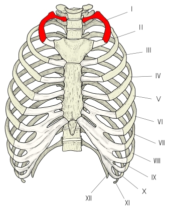
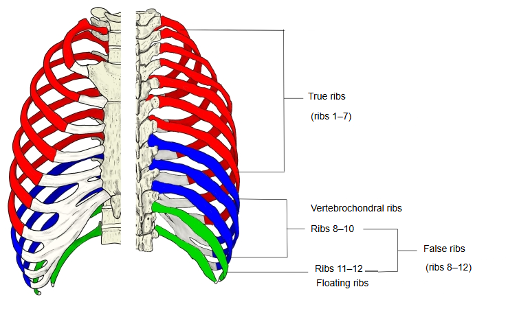
The ribs are divided into true ribs and false ribs; the first rib is the uppermost of the seven true ribs.
- True ribs : first rib, second to seventh ribs.
- False ribs
- Vertebrochondral ribs : eighth, ninth and tenth ribs.
- Floating ribs : eleventh and twelfth ribs.
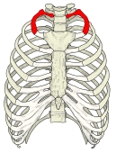 Thorax
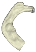 Right first rib (superior view)
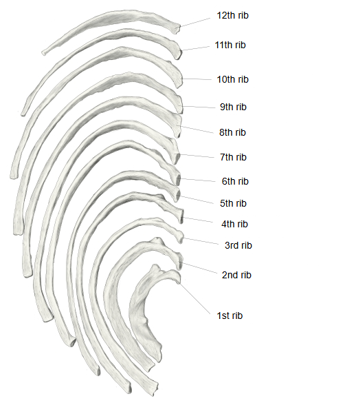
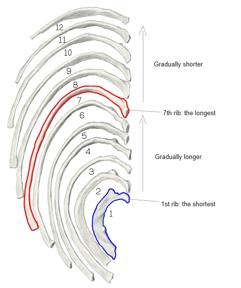
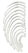 Right ribs (superior view)
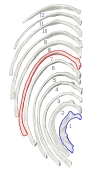 Lengths of the ribs
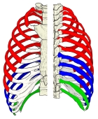 True and false ribs
Shape
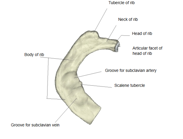
The first rib is bent strongly like a bow but is hardly twisted, so it is flat, with an upper and a lower surface. The neck is slender, but the body is wide. Telling the upper surface from the lower one is not as easy as on the other ribs.
So the first rib is one of the four ribs that differ most from the typical rib shape (the 10th rib, whose head has a single facet, is also counted as atypical in some texts).
the typical shape
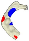
Features found only on the first rib
The following features are found on the first rib and not on the other ribs. The descriptions here are brief; see the page of each term for details.
| Name | Description | |
|---|---|---|
| 1 | Groove for subclavian vein | the groove on which the subclavian vein lies, in front of the scalene tubercle |
| 2 | Scalene tubercle (tubercle for scalenus anterior) | the insertion of scalenus anterior |
| 3 | Groove for subclavian artery | the groove on which the subclavian artery and the lowest trunk of the brachial plexus lie, behind the scalene tubercle |
Features of other ribs not found on the first rib
· Costal groove: the first rib has no costal groove.
Attachments
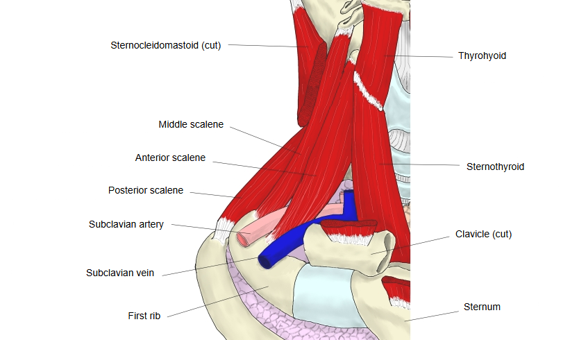
The muscles and ligaments attached to the first rib are listed below; the list may not be complete. Ligaments are divided into those attached to the bony part of the rib (costal bone) and those attached to the costal cartilage; whether ligaments of the costal cartilage should count as ligaments of the rib is open to debate.
| Name | Attachment | |||
|---|---|---|---|---|
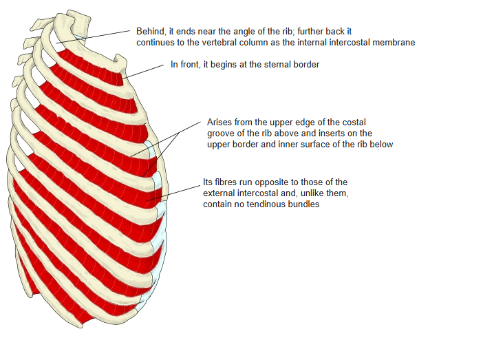
| Muscles | Origin | 1 | Internal intercostal muscle | from the upper margin of the costal groove (the first rib, which has no costal groove: from its outer border) to the upper border of the body of the rib below |
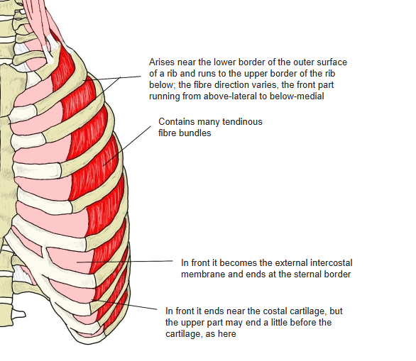
| 2 | External intercostal muscles | all ribs: from the lower border of the outer surface to the upper border of the outer surface of the rib below | ||
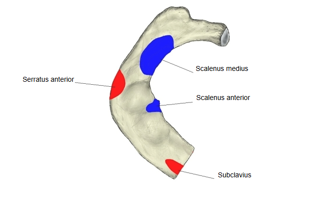
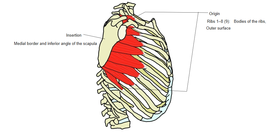
| 3 | Serratus anterior | outer surface of the bodies of the first to eighth (or ninth) ribs | ||
| 4 | Innermost intercostal muscle | not always present | ||
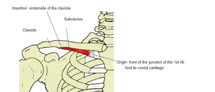
| 5 | Subclavius | first rib, near its junction with the costal cartilage ⇔ underside of the clavicle | ||
| Insertion | 6 | Scalenus anterior | first rib (scalene tubercle) ⇔ anterior tubercles of the transverse processes of C3-C6 (cervical vertebrae) | |
| 7 | Scalenus medius | first rib, behind the groove for the subclavian artery ⇔ transverse processes of C2-C7 (posterior tubercles; sources differ) | ||
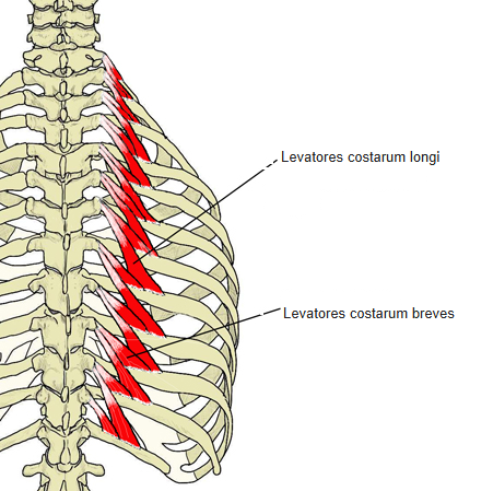
| 8 | Levatores costarum | upper border of the rib between the tubercle and the angle ⇔ transverse processes of C7-T11 (for the first rib: C7) | ||
| Ligaments | Bony rib | 1 | Radiate ligament of head of rib | head of rib ⇔ vertebral body (T1; for the first rib there is no intervertebral disc to reach) |
| 2 | Costotransverse ligament | neck of rib ⇔ transverse process | ||
| 3 | Lateral costotransverse ligament | tubercle of rib ⇔ tip of the transverse process | ||
| 4 | Costoclavicular ligament | first rib (upper border, near the costal cartilage) ⇔ clavicle (impression for costoclavicular ligament) | ||
| Costal cartilage | 1 | Radiate sternocostal ligaments | costal cartilage (front end) ⇔ sternum | |
| 2 | Intra-articular sternocostal ligament | costal cartilage (front end) ⇔ costal notch of the sternum; usually absent for the first rib, whose cartilage joins the manubrium directly |
 Origins and insertions
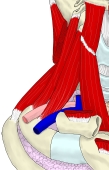 Scalene muscles
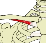 Subclavius
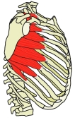 Serratus anterior
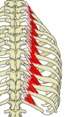 Levatores costarum
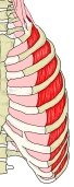 External intercostal muscles
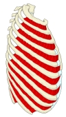 Internal intercostal muscles
Reference websites
[Wikipedia]
· Rib, Wikipedia.
· Rib cage, Wikipedia.
[Websites with illustrations]
· Ribs: anatomy, ligaments and clinical notes (Kenhub)
· The ribs (typical and atypical ribs) (TeachMeAnatomy)
· Video: Ribs - typical and atypical, true and false ribs (the first rib is explained from about 5:40).
Related terms
- True ribs
- Scalene tubercle
- Groove for subclavian artery
- Groove for subclavian vein
- Head of rib
- Costal groove
- Ribs