Frontal process (Latin: processus frontalis)
An upward projection that reaches the frontal bone; the zygomatic bone and the maxilla each have one. Both help to frame the opening of the orbit: the zygomatic one at its lateral margin, the maxillary one beside the nose at its medial margin.
Frontal process of the zygomatic bone
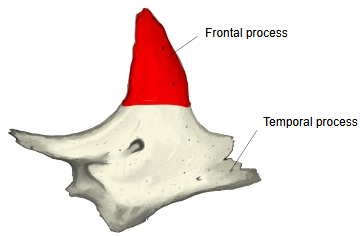
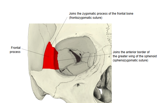
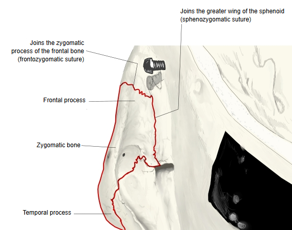
One of the two processes of the zygomatic bone. It runs upwards and joins the zygomatic process of the frontal bone and the front border of the greater wing of the sphenoid.
zygomatic bone
- Frontal process
- Frontal bone (zygomatic process) : joint: frontozygomatic suture
- Sphenoid (front border of the greater wing) : joint: sphenozygomatic suture
- Temporal process
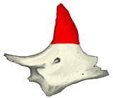 Zygomatic bone
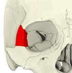 Zygomatic bone: frontal process at the orbit
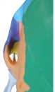 Zygomatic bone: from behind
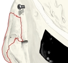 Zygomatic bone: sutures of the frontal process
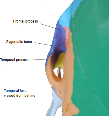
Its front edge forms the lateral margin of the orbit; its back edge borders the temporal fossa and gives attachment to the temporal fascia. A small marginal tubercle may be felt on this back edge.
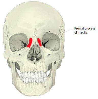
Frontal process of the maxilla
· The zygomatic bone also has a process of the same name (see above).
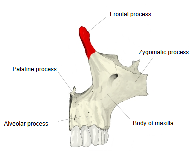
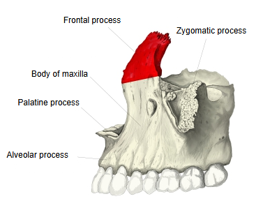
- Body of maxilla : the main, roughly pyramid-shaped part of the maxilla; it contains the maxillary sinus and has four surfaces.
- Processes
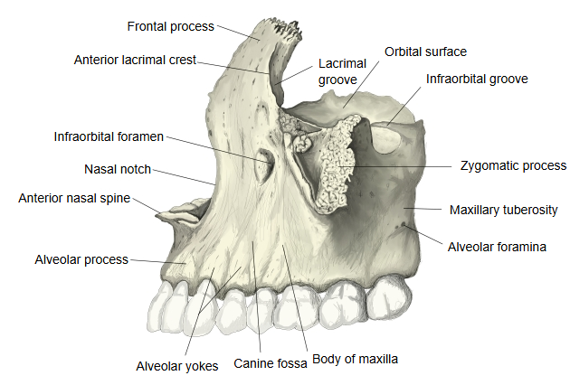
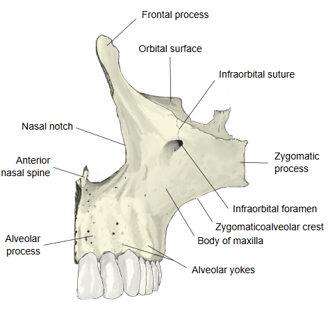
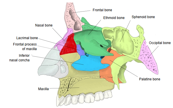
- Frontal process : a long, flat process reaching upwards, between the nasal bone and the lacrimal bone.
- Zygomatic process : a thick, short process from the lateral side of the body that joins the zygomatic bone.
- Palatine process : projects almost horizontally inwards from the lower part of the body.
- Alveolar process : the downward continuation of the body, holding the sockets for the roots of the teeth.
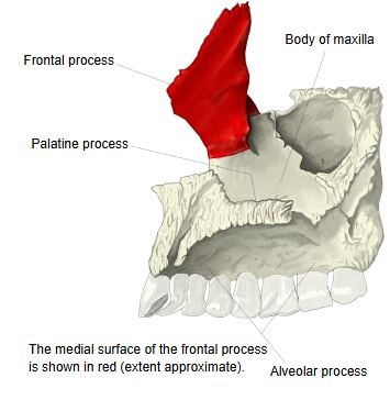
The frontal process has two surfaces: medial and lateral.
of the maxilla
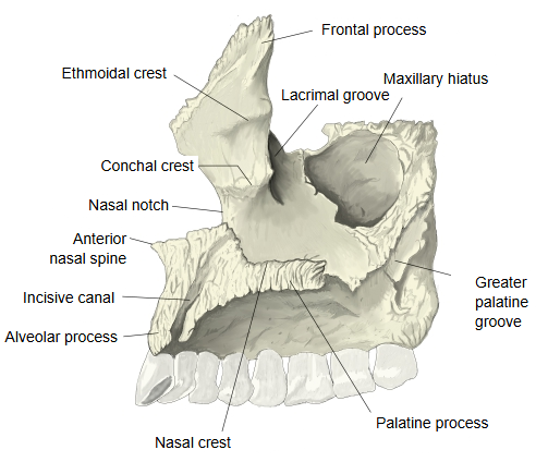
- Medial surface : faces the nasal cavity (nasal surface) and forms part of its lateral wall.
- Ethmoidal crest : the upper of two crests; attachment of the middle nasal concha.
- Conchal crest : the lower crest; attachment of the inferior nasal concha.
- Lateral surface : smooth, continuing the anterior surface of the body.
- Anterior lacrimal crest : with the posterior lacrimal crest of the lacrimal bone, it bounds the fossa for the lacrimal sac.
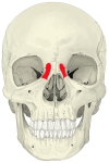 Maxilla: skull (anterior view)
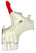 Maxilla: anterior view
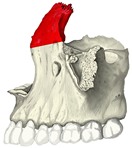 Maxilla: lateral view
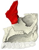 Maxilla: medial view
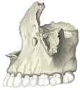 Maxilla: lateral view, main features
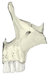 Maxilla: anterior view, main features
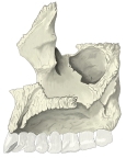 Maxilla: medial view, main features
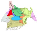 Lateral wall of nasal cavity
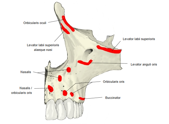
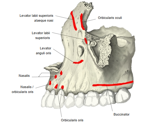
Attached muscles and ligaments
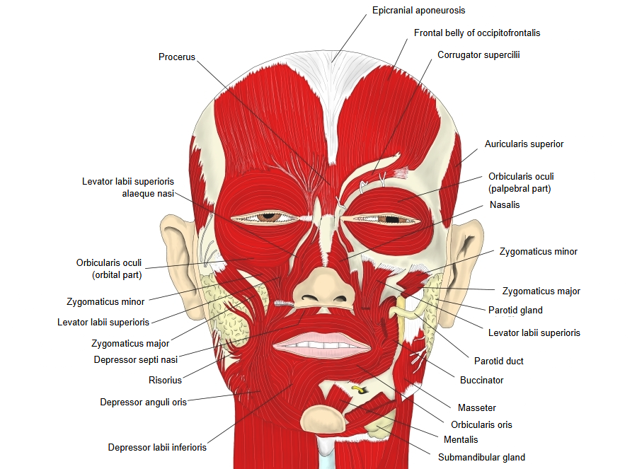
· Levator labii superioris alaeque nasi: runs from the frontal process to the skin of the upper lip and the wing of the nose.
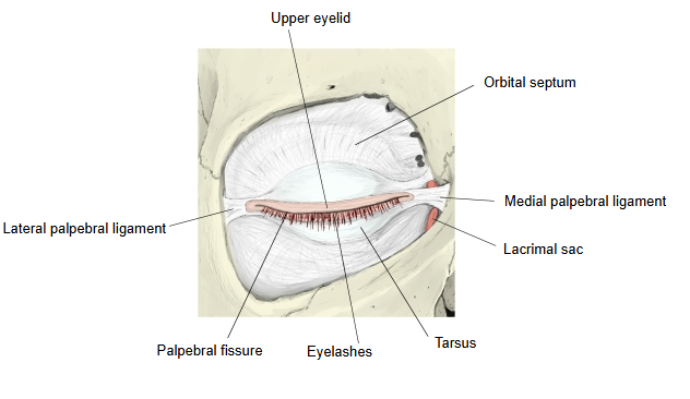
· Orbicularis oculi: the muscle around the palpebral fissure.
· Medial palpebral ligament: at the medial angle of the eye, between the frontal process and the tarsal plates.
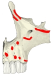 Maxilla: muscle attachments (anterior)
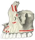 Maxilla: muscle attachments (lateral)
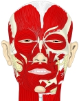 Muscles of facial expression
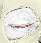 Eyelids and palpebral ligaments
The frontal process is a strong plate that projects upwards, medially and backwards from the body of the maxilla, between the nasal bone in front and the lacrimal bone behind. Its upper border joins the frontal bone; its front border is thin and serrated and joins the nasal bone; its back border is thick and hollowed into a vertical groove, the lacrimal groove, which with the lacrimal bone forms the fossa for the lacrimal sac.
Its lateral surface is smooth and gives attachment to the muscles and ligament listed above. Its medial surface forms part of the lateral wall of the nasal cavity; below the ethmoidal crest it bounds part of the middle nasal meatus, and below the conchal crest part of the inferior nasal meatus.
⇒ Back to the zygomatic bone page
Where it is found
Related terms
- Body of maxilla
- Zygomatic process
- Temporal process
- Frontozygomatic suture
- Zygomaticomaxillary suture
- Lacrimal groove
- Nasal bone
References and sources
- Further reading: Frontal process of maxilla, Wikipedia.
- Further reading: Zygomatic bone, Wikipedia.
- Federative International Programme for Anatomical Terminology (FIPAT). Terminologia Anatomica, 2nd ed. (TA2), 2019.