Latin: lingua
Overview
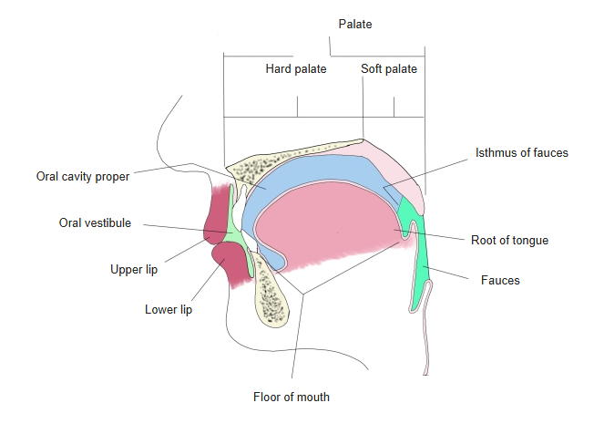
- A muscular organ covered by mucous membrane, lying in the floor of the oral cavity and reaching back into the oral part of the pharynx.
- Moves food during chewing and swallowing, carries most of the taste buds, and shapes sounds in speech.
- Anchored by its muscles to the hyoid bone, the mandible, the styloid process, the soft palate and the pharyngeal wall.
- Has a rich supply of blood vessels and nerves.
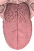
Parts of the tongue 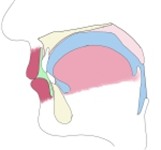
Oral cavity (schematic section)
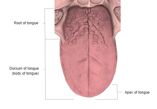
Parts and surfaces
| Part | Description |
|---|---|
| Apex of tongue | The free tip at the front |
| Body of tongue | The front two-thirds (oral part); its upper surface is the dorsum of tongue |
| Root of tongue | The back third (pharyngeal part), facing the oropharynx; fixed to the hyoid bone and mandible |
| Sulcus terminalis | V-shaped groove, pointing backwards, between the body and the root |
| Foramen cecum | Small pit at the point of the V; remnant of the embryonic thyroglossal duct |
| Median sulcus | Shallow groove along the midline of the dorsum, over the lingual septum |
| Margin of tongue | The rounded side edge of the apex and body |
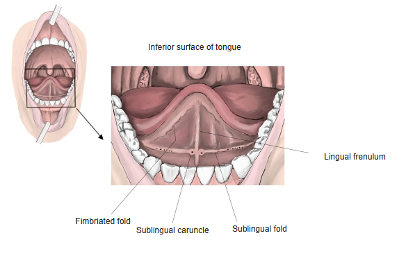
| Inferior surface | Smooth underside, joined to the floor of the mouth by the lingual frenulum |
- Beside the frenulum are the sublingual caruncles, where the ducts of the submandibular gland and the major duct of the sublingual gland open.
- The sublingual folds run back from the caruncles over the sublingual glands; the fringed fimbriated folds run forward towards the apex.
- Between the root and the epiglottis lie one median and two lateral glossoepiglottic folds; the hollows between them are the epiglottic valleculae.
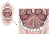
Inferior surface of tongue
Lingual papillae
| Papilla | Where | Notes |
|---|---|---|
| Filiform papillae | Whole dorsum of the body | The most numerous; slender keratinised tips give the velvety, whitish surface; no taste buds |
| Fungiform papillae | Among the filiform papillae, most at the tip and sides | Mushroom-shaped red dots; carry taste buds |
| Vallate papillae | One V-shaped row of about 8–12 just in front of the sulcus terminalis | Large, each sunk in a circular trench; many taste buds in the walls; serous (von Ebner) glands open into the trench |
| Foliate papillae | Side edges at the back of the body | Parallel leaf-like folds with taste buds; best developed in children |
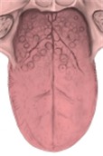
Lingual papillae
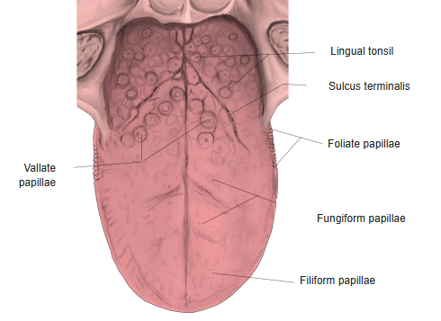
Mucosa of the root
- The mucosa of the root has no papillae; it is raised into low bumps by the lingual follicles, each a group of lymphoid nodules around a small crypt.
- Together the follicles form the lingual tonsil. It thins out towards the epiglottis behind and the palatoglossal arches at the sides.
- With the palatine and pharyngeal tonsils it forms a ring of lymphoid tissue around the entrance to the pharynx; its structure is like a small palatine tonsil.
Muscles of the tongue
| Muscle | Group | Main action |
|---|---|---|
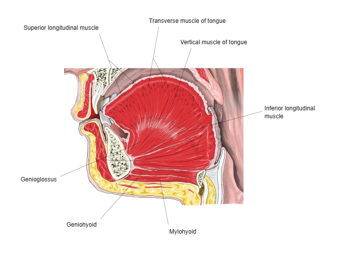
| Genioglossus | Extrinsic | Pulls the tongue forward (protrusion) and depresses its centre |
| Hyoglossus | Extrinsic | Depresses and retracts the tongue, pulling its sides down |
| Styloglossus | Extrinsic | Retracts the tongue and raises the dorsum |
| Palatoglossus | Extrinsic (also counted with the soft palate) | Raises the root and narrows the isthmus of fauces |
| Superior longitudinal muscle | Intrinsic | Shortens the tongue and turns the tip and sides up |
| Inferior longitudinal muscle | Intrinsic | Shortens the tongue and turns the tip down |
| Transverse muscle of tongue | Intrinsic | Narrows and lengthens the tongue |
| Vertical muscle of tongue | Intrinsic | Flattens and widens the tongue |
- Extrinsic muscles arise outside the tongue and change its position; intrinsic muscles lie wholly inside it and change its shape.
- All are skeletal muscles, divided into right and left halves by the fibrous lingual septum and separated into small bundles by fatty connective tissue.
- When the hyoglossus, styloglossus and genioglossus of one side act alone, the tongue bends towards that side.
- All are supplied by the hypoglossal nerve, except the palatoglossus (vagus nerve through the pharyngeal plexus).
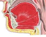
Muscles of the tongue (median section)
Blood supply, lymph and nerves
| Supply | |
|---|---|
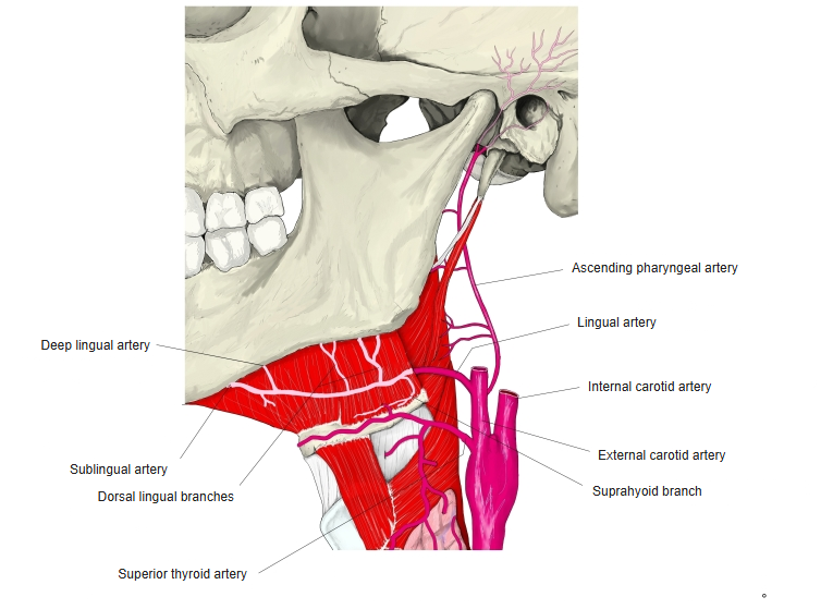
| Arteries | Lingual artery from the external carotid artery (dorsal lingual, sublingual and deep lingual branches); the root also gets tonsillar branches of the facial artery and the ascending pharyngeal artery. Right and left arteries hardly join across the midline. |
| Veins | Lingual, dorsal lingual and sublingual veins, draining to the internal jugular vein (directly or through the facial vein) |
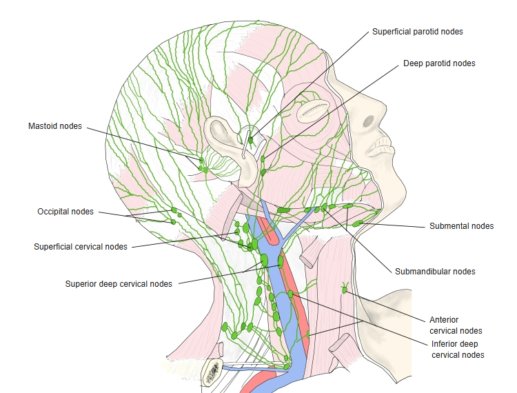
| Lymph | Tip: submental nodes; body: submandibular and deep cervical nodes; root: deep cervical nodes of both sides |
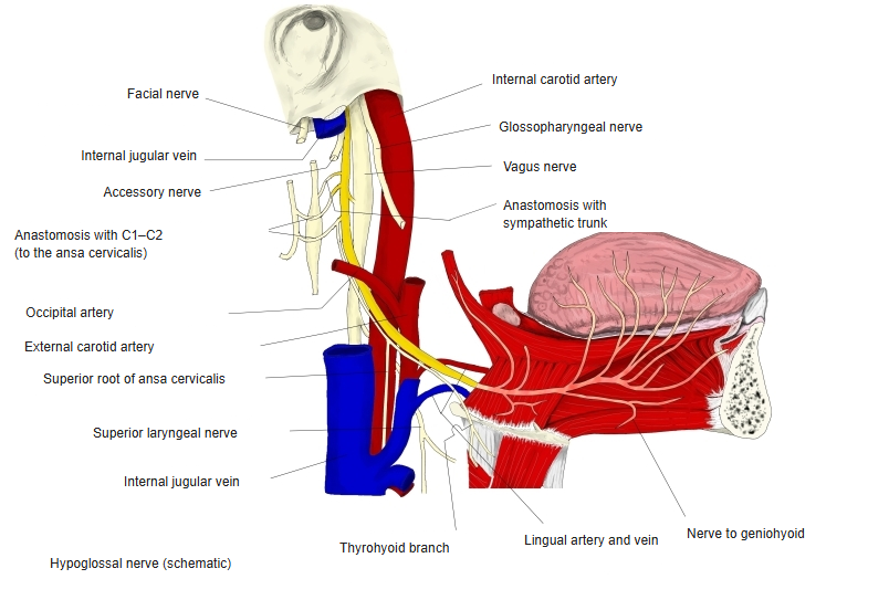
| Motor nerve | Hypoglossal nerve (palatoglossus: vagus nerve) |
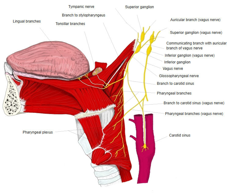
| General sensation | Front two-thirds: lingual nerve (from the mandibular nerve); back third: glossopharyngeal nerve; small area in front of the epiglottis: superior laryngeal nerve (vagus) |
| Taste | Front two-thirds: chorda tympani of the facial nerve, running with the lingual nerve; back third including the vallate papillae: glossopharyngeal nerve; epiglottic region: vagus nerve |
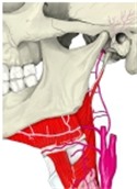
Lingual artery 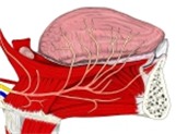
Hypoglossal nerve 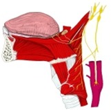
Glossopharyngeal and vagus nerves 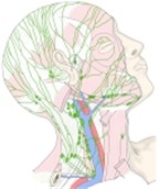
Lymph nodes of the head and neck
Function
- Chewing: keeps food between the teeth and mixes it with saliva.
- Swallowing: presses the bolus up against the palate and pushes it back into the pharynx.
- Taste: the taste buds of the fungiform, vallate and foliate papillae.
- Speech: changes shape and position quickly to form many speech sounds.
Clinical notes
- Tongue-tie (ankyloglossia): a short lingual frenulum limits movement of the tip.
- Hypoglossal nerve injury: the protruded tongue deviates towards the paralysed side.
- Thyroglossal duct cyst: can form anywhere along the path from the foramen cecum down to the thyroid gland.
- Sublingual drugs: the thin mucosa of the inferior surface allows quick absorption of some medicines.
References and sources
- Further reading: Tongue, Wikipedia.